Contact Person:Dr. Safwan Sayyal
Working Hours:Mon - Fri: 9:00 AM - 5:00 PM
Contact:info@sayyalhealth.com




Published: August 31st, 2026Last updated: September 23rd, 2026
The stages of melanoma describe how far the cancer has grown or spread when it is diagnosed. Melanoma is staged from Stage 0, when abnormal melanocytes are confined to the epidermis, through Stage 4, when the cancer has spread to distant parts of the body.
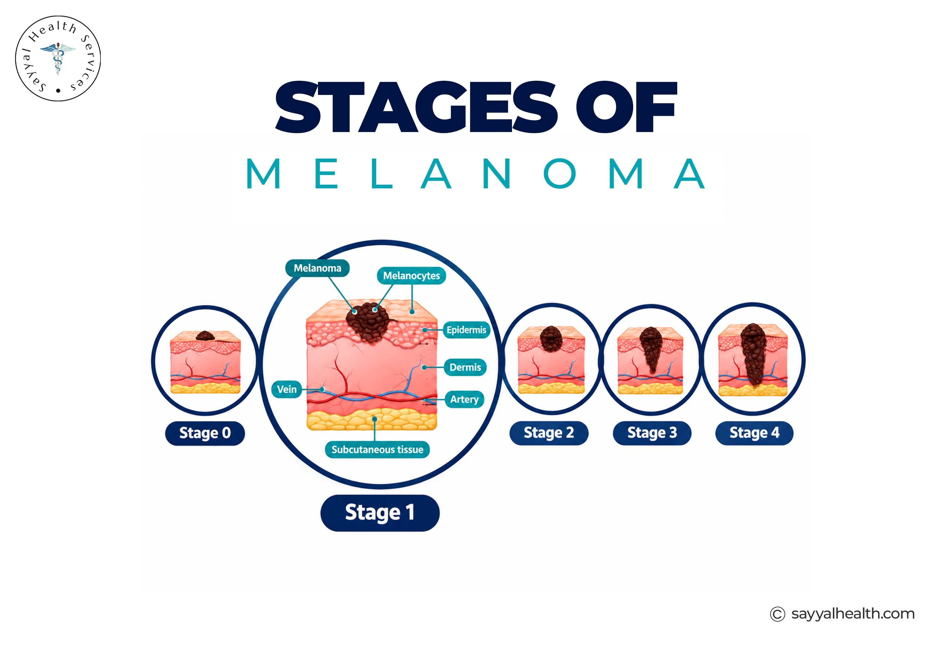
Here is a simple overview before we go section by section.
|
Stage |
General Meaning |
|
Stage 0 |
Melanoma in situ, confined to the epidermis |
|
Stage 1 |
Localised invasive melanoma with no detected spread to lymph nodes or distant sites |
|
Stage 2 |
Localised melanoma that is thicker and/or ulcerated, without detected spread to lymph nodes or distant sites |
|
Stage 3 |
Melanoma with regional spread to nearby lymph nodes and/or nearby skin or lymphatic tissue |
|
Stage 4 |
Melanoma has spread to distant parts of the body |
Reminder: Doctors assign the exact stage using detailed pathology results, not just a general description.
Melanoma staging is the process doctors use to describe how advanced a melanoma is at diagnosis. It matters for two practical reasons:
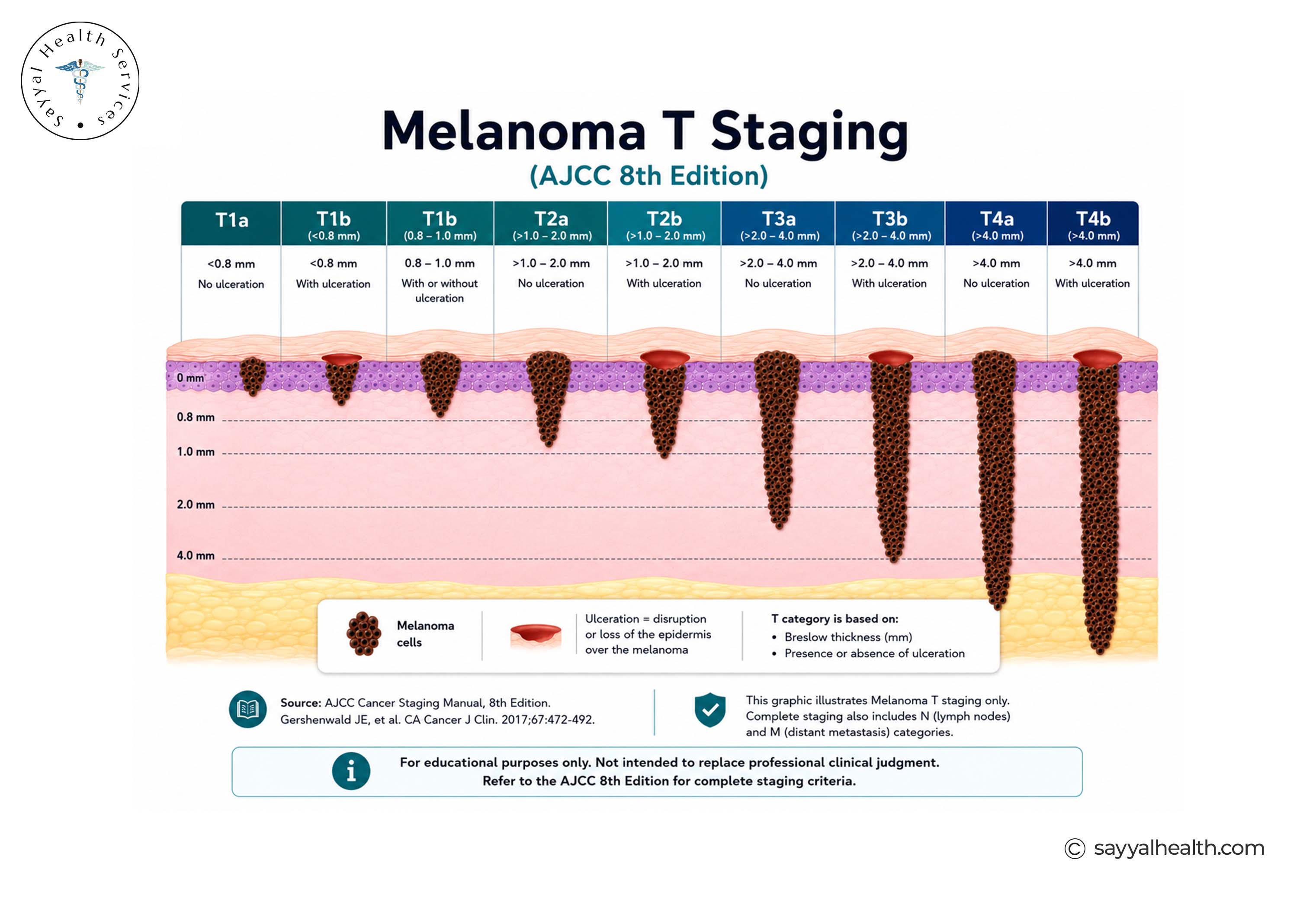
Melanoma is staged using the TNM system (Tumour, Nodes and Metastasis), developed by the American Joint Committee on Cancer (AJCC) and used internationally. TNM describes three key features: the primary tumour, whether the melanoma has spread to nearby lymph nodes or regional tissues, and whether it has spread to distant parts of the body.
The T category describes the primary melanoma itself, mainly its thickness (Breslow thickness) and whether the surface has ulcerated. Thicker and ulcerated melanomas generally carry a higher T category.
The N category describes whether melanoma cells have reached nearby lymph nodes, and if so, how many nodes are involved and whether the spread is detectable by touch/imaging or only on pathology testing.
The M category describes whether melanoma has spread to distant sites, parts of the body away from the original mole or lesion, such as internal organs, distant skin, or distant lymph nodes.
A pathologist and treating doctor combine the T, N and M findings to arrive at an overall stage from 0 to 4, with several stages broken into sub-stages (like 1A, 1B, or 3A–3D) that reflect finer differences in prognosis.
Breslow thickness measures the depth of an invasive melanoma in millimetres, from the top of the epidermis to the deepest point of tumour invasion. It is one of the key factors used to determine the T category in melanoma staging.
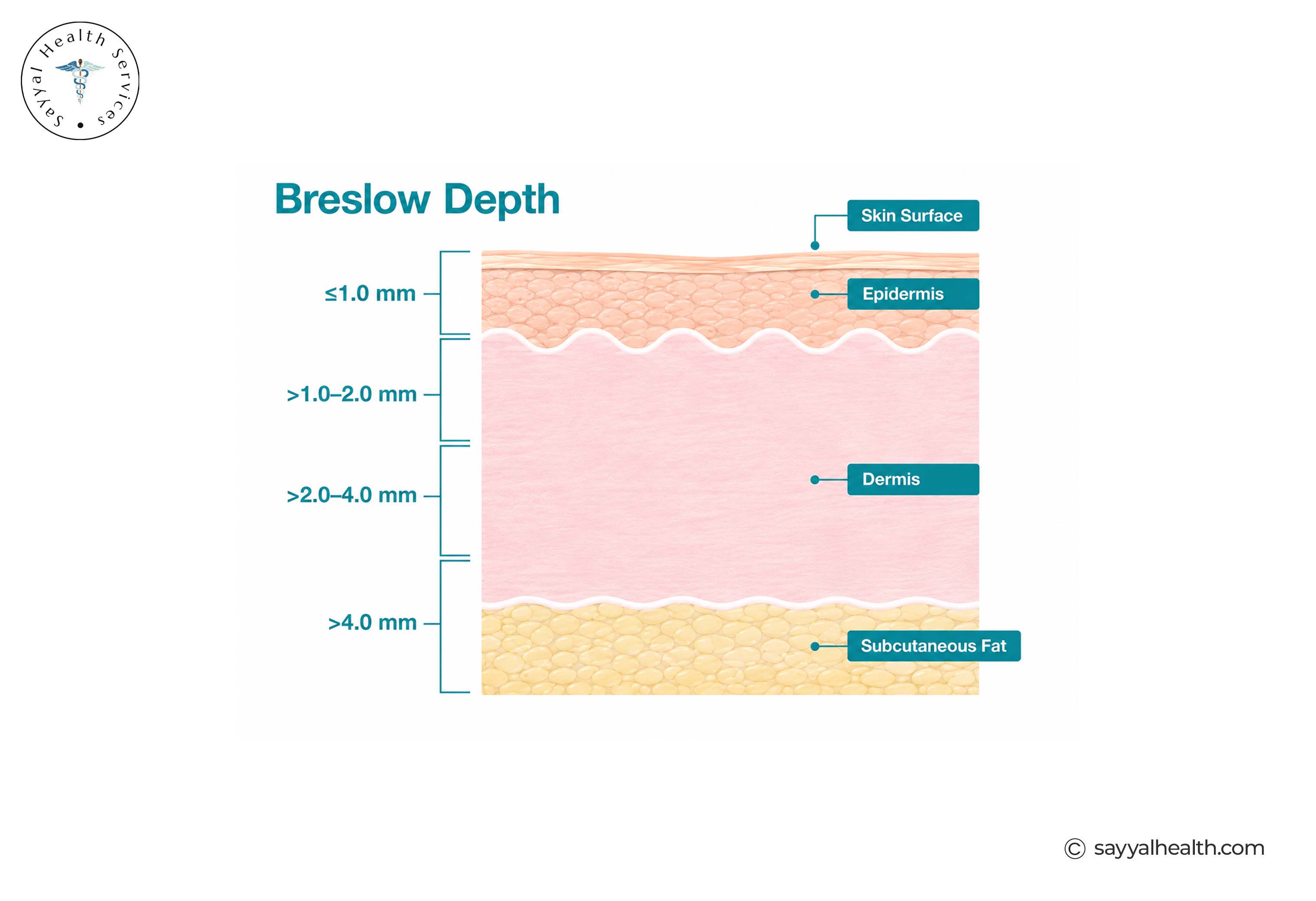
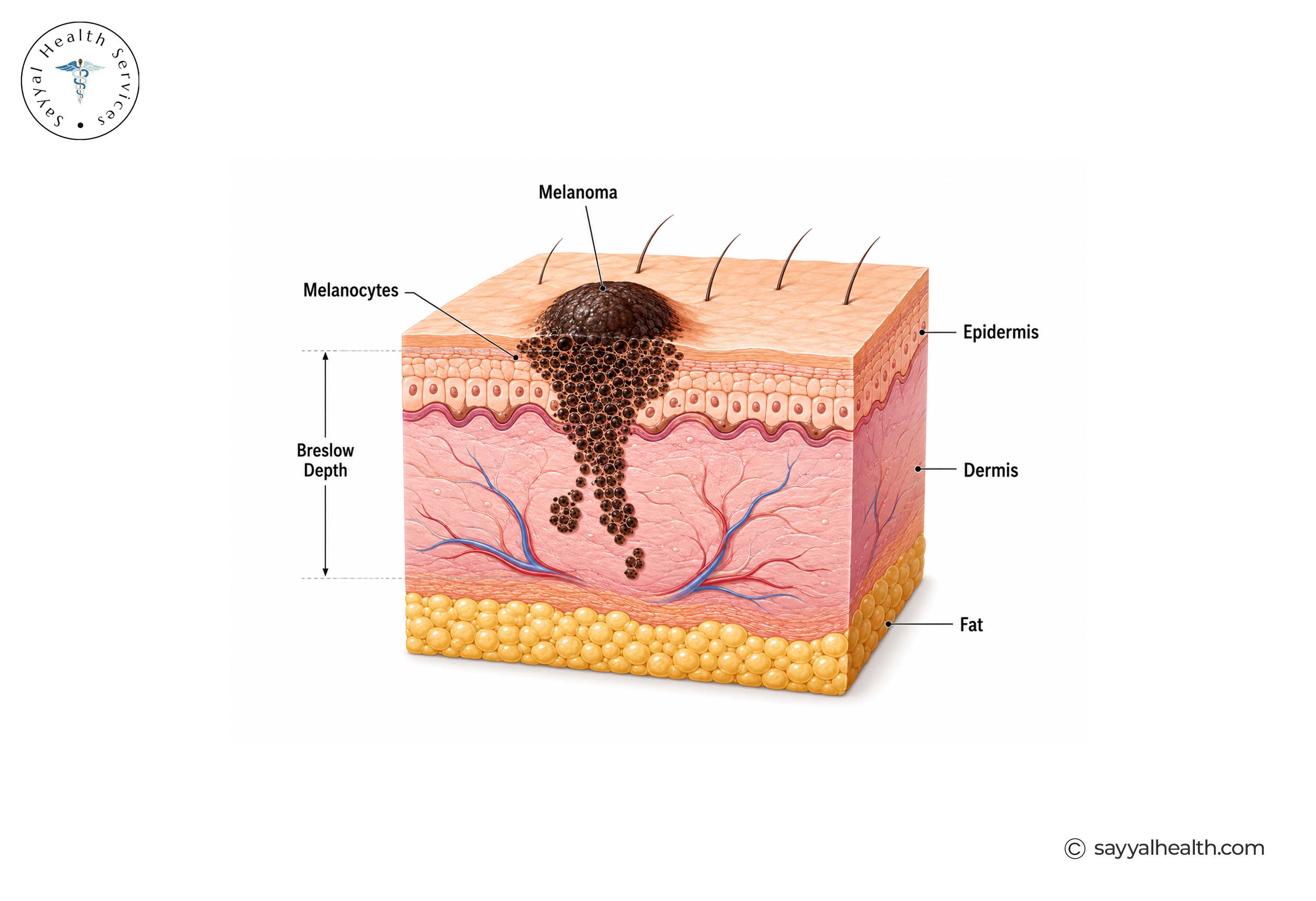
A pathologist determines Breslow thickness by examining the biopsy specimen under a microscope and measuring the depth of the invasive melanoma in millimetres.
Breslow thickness feeds directly into the T category of staging. In the AJCC 8th edition staging system, Breslow thickness categories are divided at key thresholds of 0.8 mm, 1.0 mm, 2.0 mm and 4.0 mm, together with whether the melanoma is ulcerated.
As a general rule, the thicker the melanoma, the more it typically influences the overall stage and the treatment plan a doctor recommends, though thickness is only one part of the picture, alongside ulceration and, at more advanced stages, lymph node and metastasis findings.
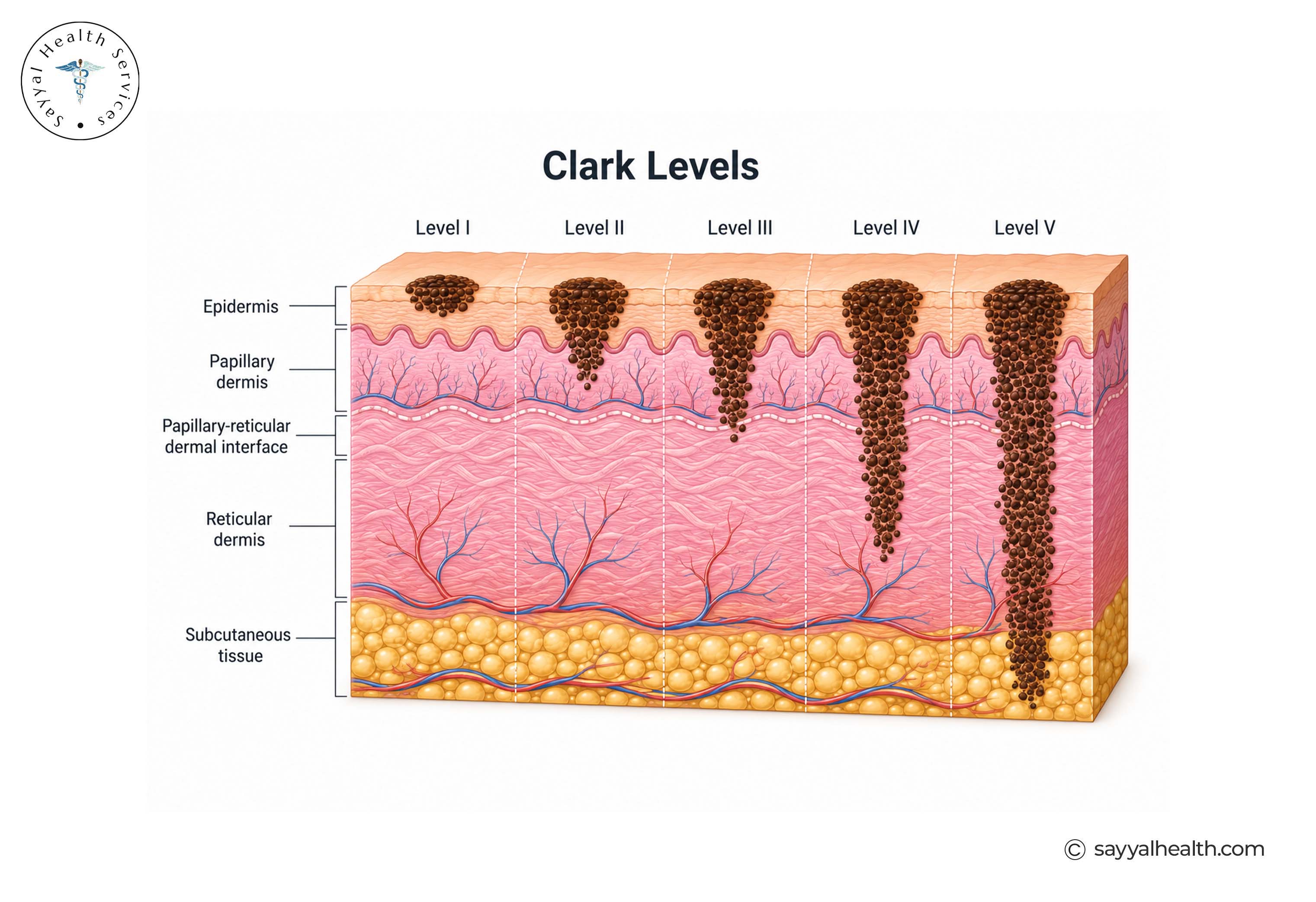
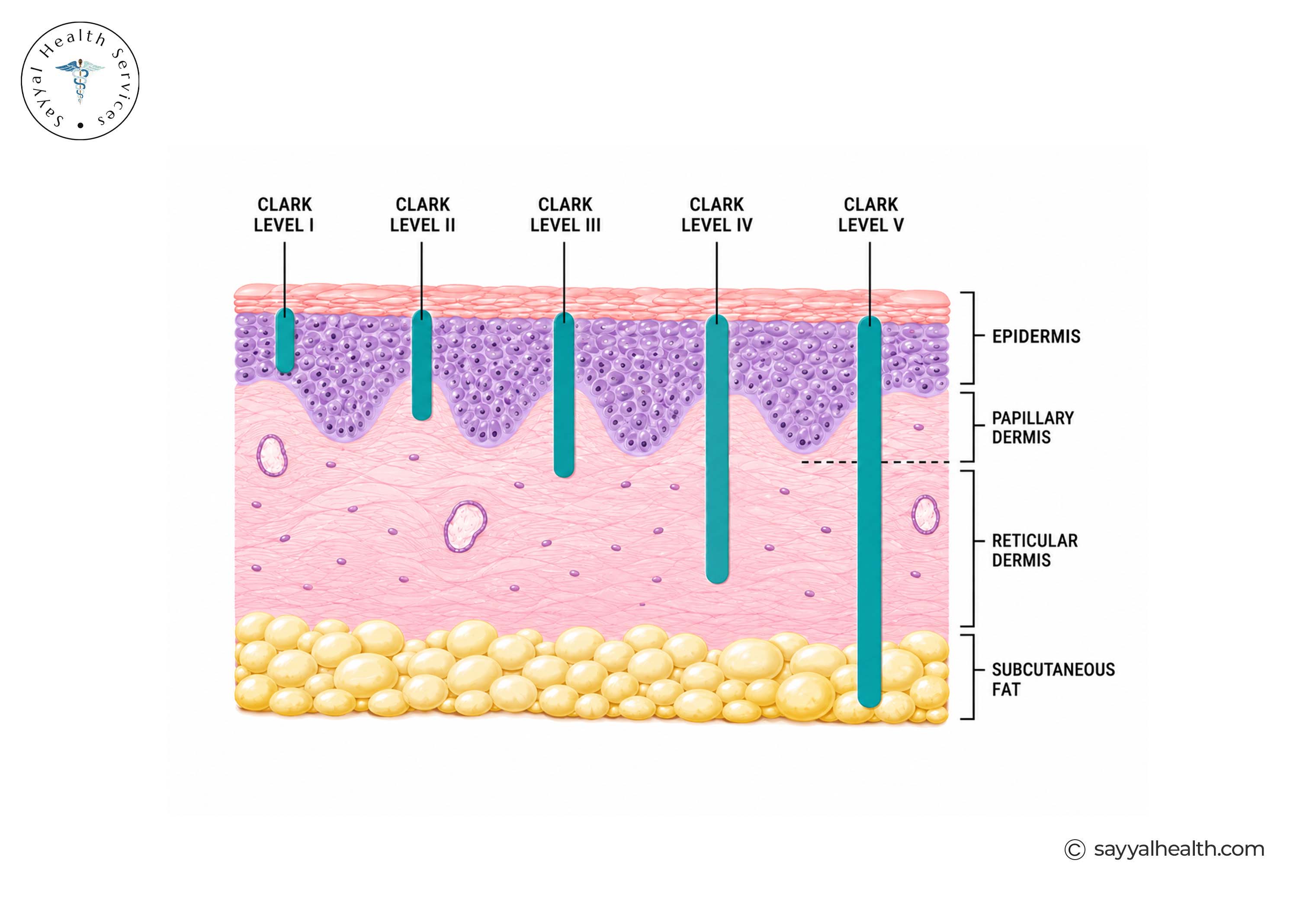
Clark level is an older method of describing how deeply a melanoma has invaded the skin based on the anatomical layer reached. Breslow thickness is now the more important measurement used in modern melanoma staging.
Stage 0 melanoma means the abnormal melanocytes (pigment-producing cells) are confined entirely to the epidermis, the outermost layer of skin. This is also called melanoma in situ, "in situ" simply means "in its original place," because the abnormal cells haven't broken through into the deeper layer of skin (the dermis).
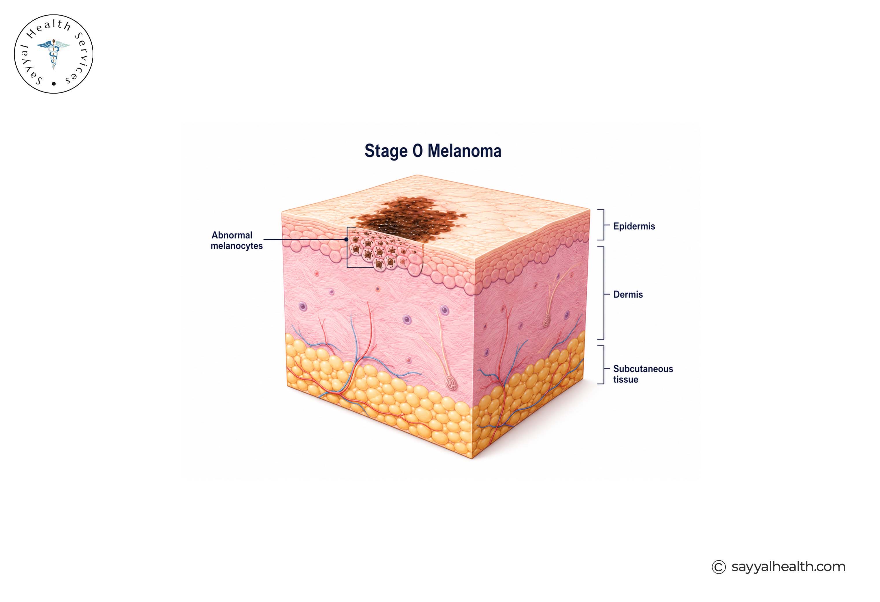
It's important not to think of Stage 0 as harmless or "not really cancer." Melanoma in situ is still melanoma, but the abnormal cells remain confined to the epidermis and have not invaded the deeper dermis. Because it has not become invasive, Stage 0 melanoma is generally treated with surgical removal, with the specific approach determined by the treating clinician.
Stage 1 melanoma is localised invasive melanoma: the cancer has grown into the dermis, but it's thin, there's no evidence it has spread to lymph nodes or elsewhere, and the outlook at this stage is generally very good.
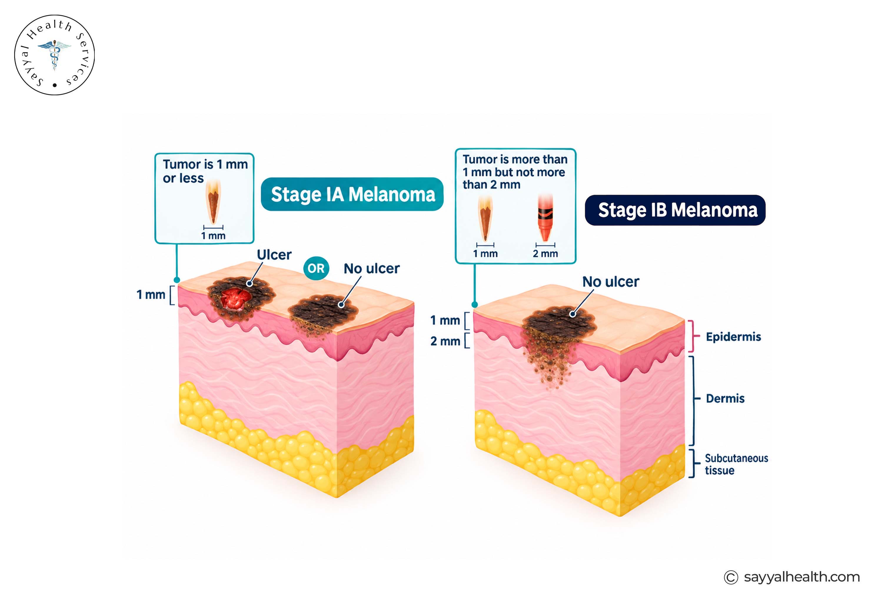
Stage 1A includes melanoma that is less than 0.8 mm thick without ulceration, with no evidence of regional or distant spread.
Stage 1B includes melanomas less than 0.8 mm thick with ulceration, or melanomas 0.8–1.0 mm thick with or without ulceration, with no evidence of regional or distant spread.
Stage 2 melanoma is still localised, which means there's no detected spread to lymph nodes or distant sites. But the tumour is thicker and/or ulcerated compared with Stage 1, which raises its risk profile.
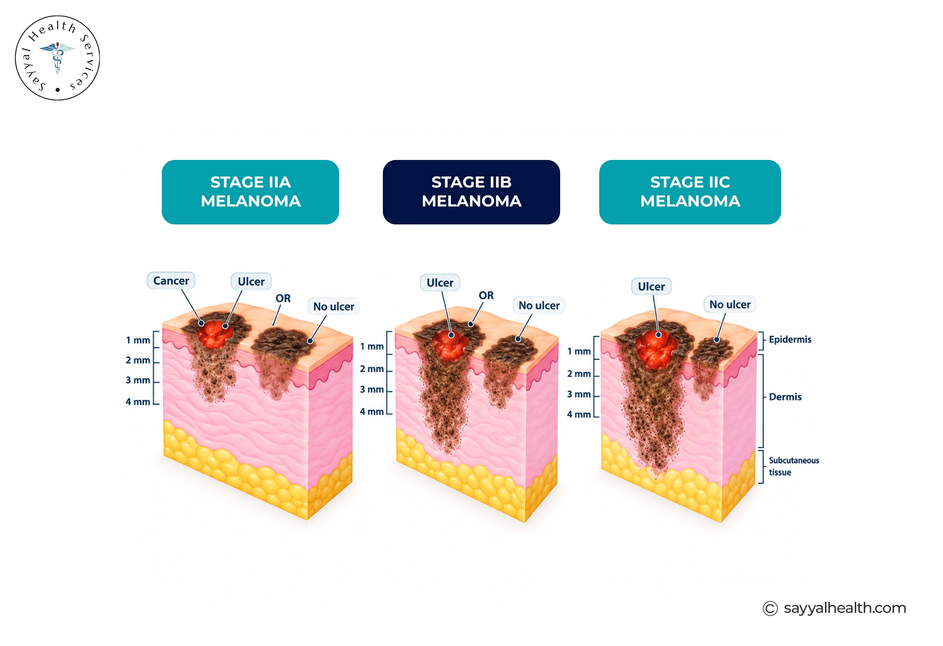
Stage 2A includes either a melanoma more than 1.0 mm but no more than 2.0 mm thick with ulceration, or a melanoma more than 2.0 mm but no more than 4.0 mm thick without ulceration.
Stage 2B includes a melanoma more than 2.0 mm but no more than 4.0 mm thick with ulceration, or a melanoma more than 4.0 mm thick without ulceration.
Stage 2C describes melanoma more than 4.0 mm thick with ulceration, without detected spread to regional lymph nodes or distant sites.
Although Stage 2C remains localised, it carries a higher risk of recurrence than earlier Stage 2 sub-stages. Depending on the individual melanoma and clinical circumstances, doctors may discuss additional evaluation, including sentinel lymph node biopsy
Stage 3 melanoma generally means the cancer has spread beyond the original site to nearby lymph nodes or nearby skin and tissue, what doctors call regional spread. This is a broad stage that covers a wide range of situations, from a single microscopic deposit in one lymph node through to more extensive regional disease.
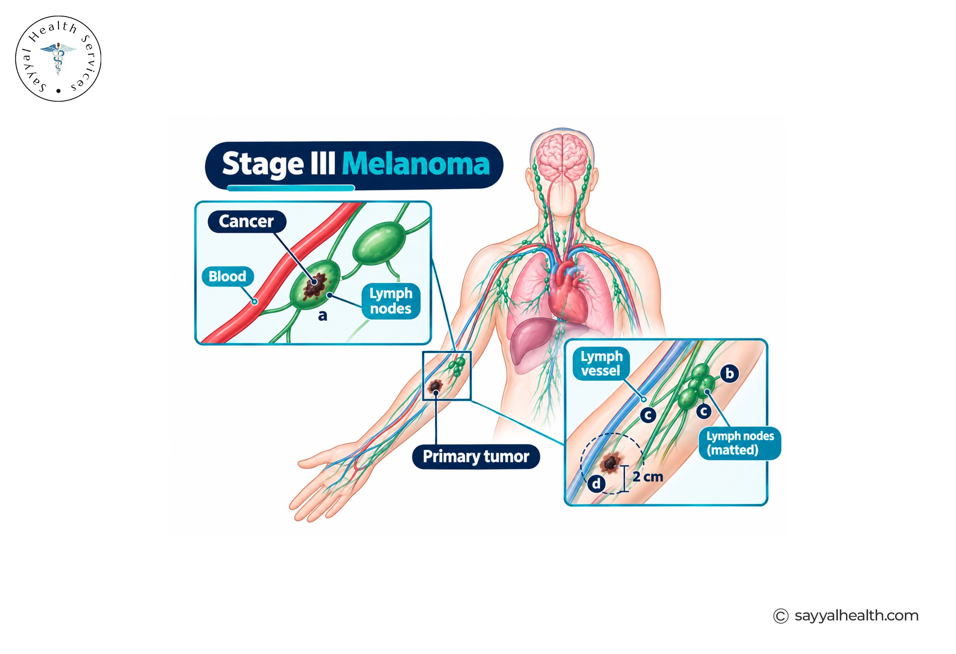
Doctors look at several things when staging Stage 3 melanoma:
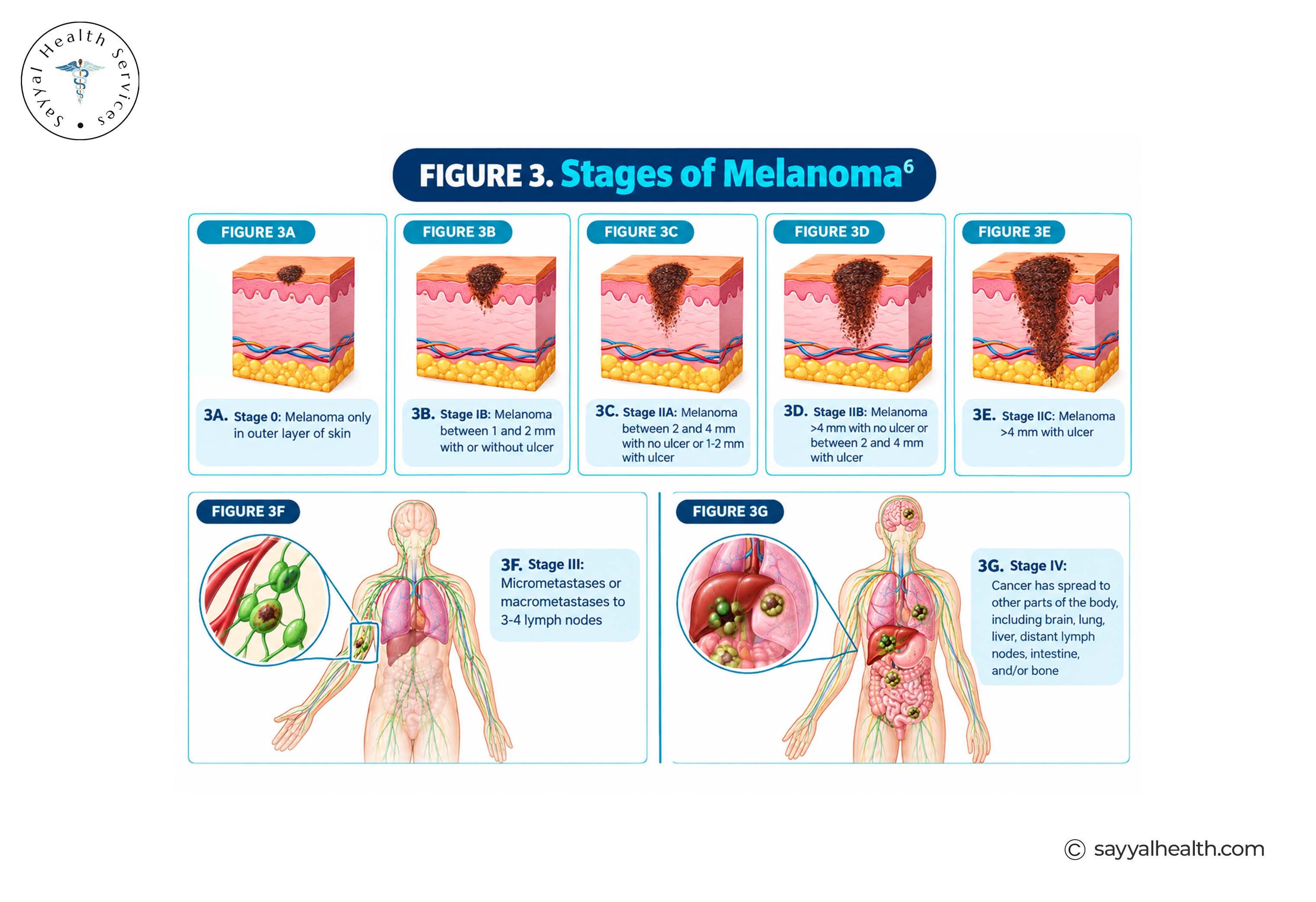
Stage 3A generally involves melanoma with limited regional spread that is detected microscopically, such as certain cases involving one or more nearby lymph nodes. The exact Stage 3A criteria depend on the primary tumour and the pattern and extent of regional metastasis.
Stage 3B includes several combinations of the primary tumour characteristics and regional spread, including certain cases involving nearby lymph nodes or satellite, in-transit or microsatellite metastases.
Stage 3C represents more extensive regional disease than Stage 3A or 3B and can involve combinations of primary tumour characteristics, lymph-node involvement and satellite, in-transit or microsatellite metastases.
Stage 3D is the highest Stage 3 sub-stage in the AJCC 8th edition system and represents extensive regional disease involving specific combinations of tumour, lymph-node and/or satellite or in-transit metastasis features.
The outlook for Stage 4 melanoma varies considerably between individuals. Factors include where and how extensively the melanoma has spread, tumour characteristics, available treatment options, overall health and how the melanoma responds to treatment.
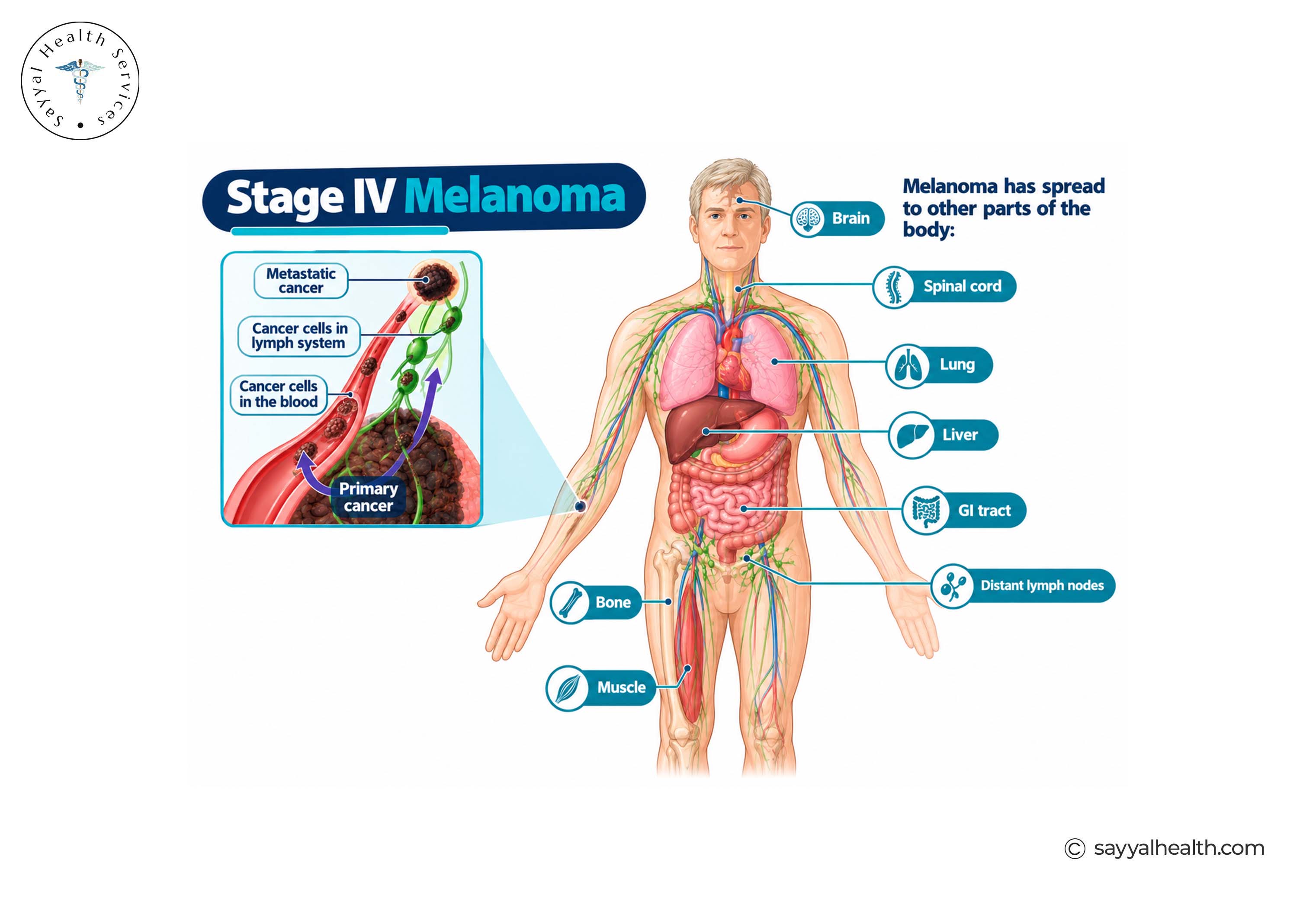
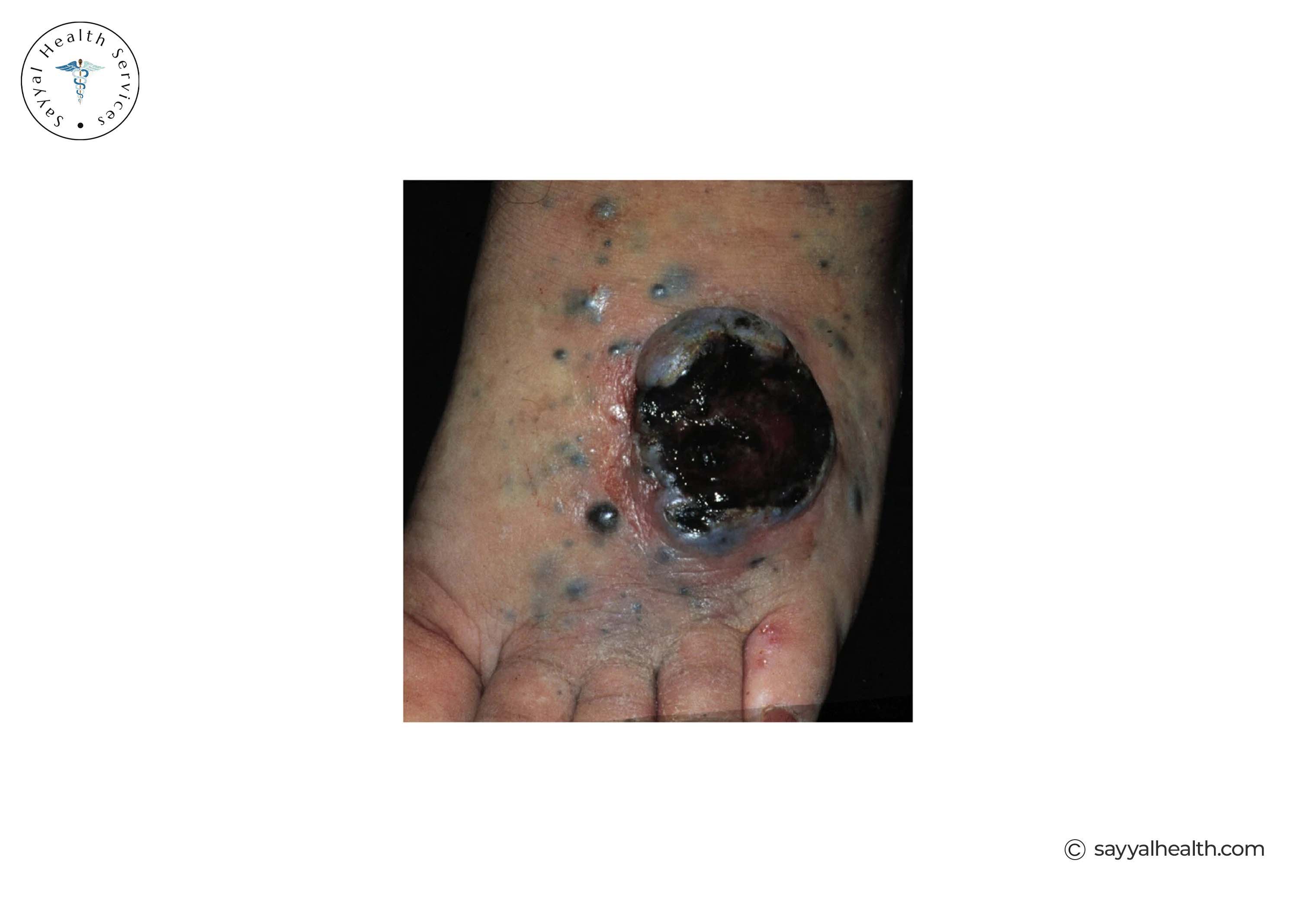
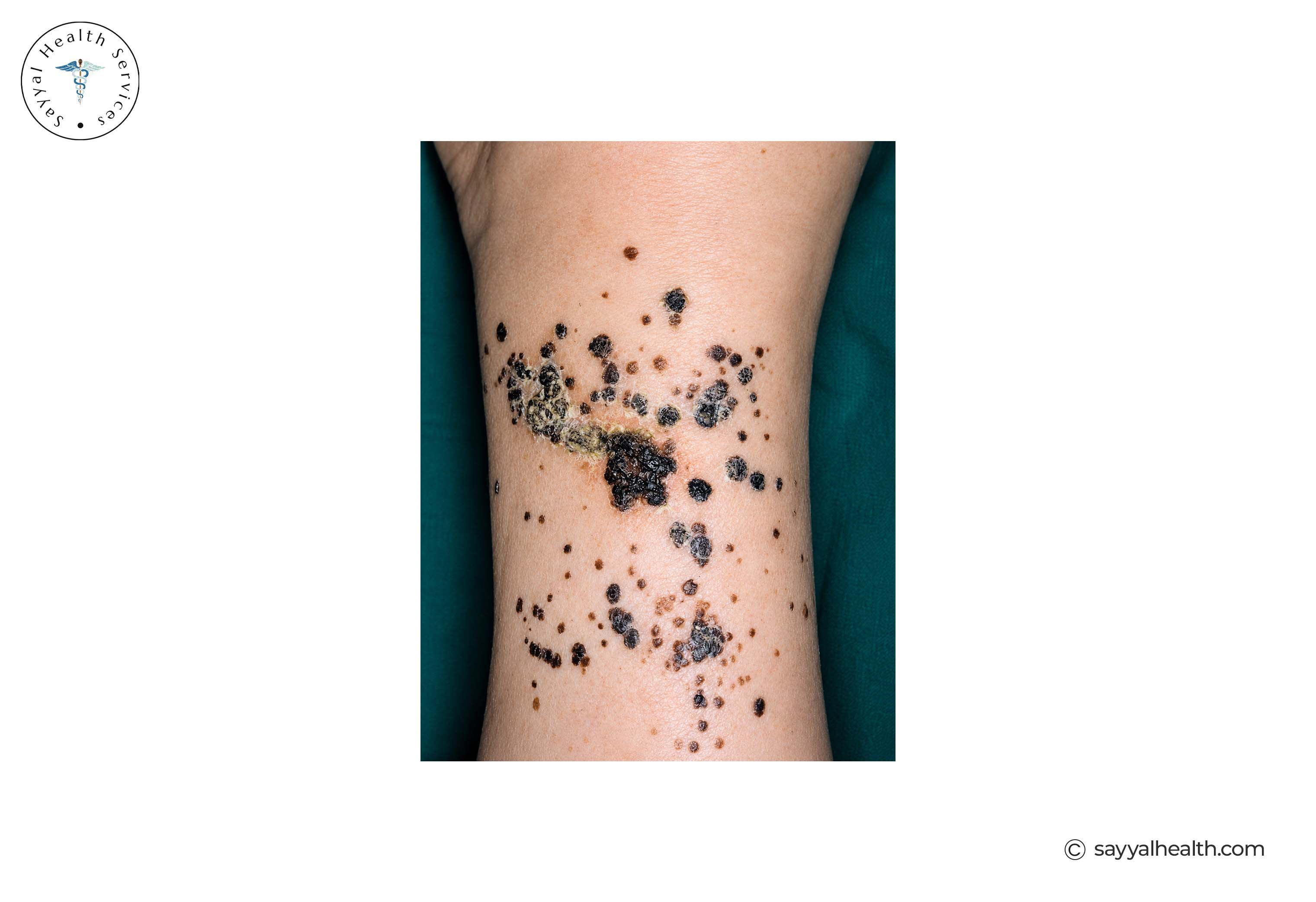
Melanoma can spread to a range of sites, including:
A Stage 4 diagnosis is understandably frightening, but it doesn't have one fixed outcome. Where the melanoma has spread, how much it has spread, a person's general health, and increasingly how well the melanoma responds to modern treatments such as immunotherapy and targeted therapy, all shape what happens next. Treatment for Stage 4 melanoma has changed substantially over the past decade, and outcomes for many patients have improved as a result.
There's no single timeline for how quickly melanoma spreads; it depends on the individual tumour, and doctors can't reliably predict an individual melanoma's behaviour just by looking at it.
That said, several factors are linked to a higher likelihood of faster or more extensive spread:
Because of this variability, the safest approach is simply not to wait and watch a changing lesion; get it assessed.
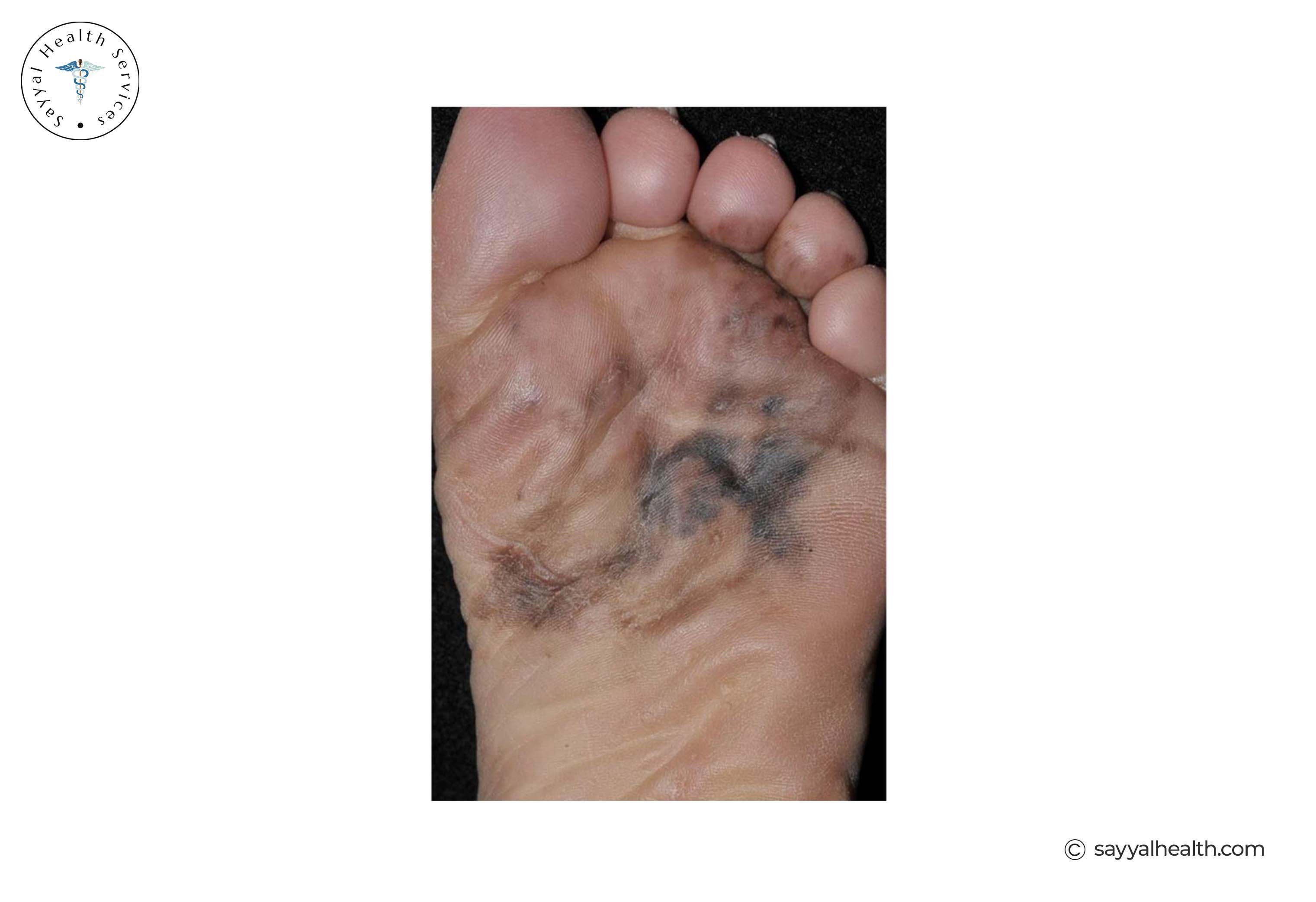
Melanoma can be flat, raised, or partly flat and partly raised; there's no single shape that defines it. Many early melanomas start flat and become raised as they grow, but this isn't a reliable rule, and some melanomas are raised from the outset.
Because appearance alone can't confirm whether a lesion is melanoma or say anything about its stage, doctors and skin-checking guidelines focus on patterns of change rather than a single visual feature. Signs worth having checked include:
No single one of these automatically means melanoma, and plenty of harmless spots share some of these features. That's exactly why a professional opinion matters more than trying to self-diagnose from appearance.
No, a photo can help flag a suspicious lesion, but it can not determine a melanoma's stage. Those are two different jobs, and it's worth keeping them separate.
Spotting a suspicious lesion is about noticing a spot that looks different from your others, or one that's changing something a photo, a skin-check app, or your own eyes can genuinely help with.
Staging a confirmed melanoma requires a biopsy and pathology assessment (to measure Breslow thickness and check for ulceration), and often further investigations such as a sentinel lymph node biopsy or imaging, depending on what the pathology shows. None of that can be done from an image.
Survival statistics for Stage 3 melanoma vary considerably depending on the exact sub-stage (3A through 3D), because Stage 3 covers such a wide range of disease extent. As a general pattern, Stage 3A carries a notably better outlook than Stage 3D, reflecting the difference between limited, microscopic lymph node involvement and more extensive regional disease.
A few things are worth keeping in mind whenever you read melanoma survival statistics:
Melanoma Institute Australia notes that overall survival for advanced melanoma has improved substantially over roughly the past 15 years, largely due to newer treatments a trend that applies broadly across regional and distant-stage disease, not just Stage 4.
If your doctor has given you a stage and sub-stage, ask them directly about the outlook for your specific situation; general statistics can't account for your individual pathology results, treatment plan or response to treatment.
Stage 4 melanoma survival has changed significantly over the past decade, with advances in treatments such as immunotherapy and targeted therapy.
According to the Australian Institute of Health and Welfare (AIHW), the 5-year relative survival rate for Australians diagnosed with Stage 4 melanoma was 26%, based on national data from 2011. However, treatments have advanced since then, so this figure should not be used to predict an individual’s outcome.
Outcomes vary depending on factors such as:
These are population-level statistics, not a personal forecast. An oncologist can provide a more meaningful assessment based on an individual's specific situation.
There is no single life-expectancy figure that applies to everyone with Stage 4 melanoma. Some people respond well to modern treatments and live for years, while others may have a more difficult course.
Survival can vary considerably depending on factors such as how far the melanoma has spread, tumour biology, the treatments available, and how well the cancer responds to treatment.
Treatment options have also changed significantly. Immunotherapy and targeted therapies have improved outcomes for many people with Stage 4 melanoma, while clinical trials continue to provide new treatment possibilities.
If you or someone you love has been diagnosed with Stage 4 melanoma, talk with an oncologist about your individual situation rather than relying on a general survival statistic.
Not all stages of skin cancer are worked out the same way. Melanoma staging relies heavily on Breslow thickness, ulceration, lymph node status and distant spread. Basal cell carcinoma and squamous cell carcinoma are the most common types of non-melanoma skin cancer. They are usually lower risk and are often assessed based on factors such as their size and location, rather than requiring the detailed staging tests commonly used for melanoma.
Go to a doctor or dermatologist to check your spot if you notice these:
None of these signs confirms melanoma on their own. Plenty of harmless spots change slightly over time, and plenty of ordinary moles itch occasionally. But a professional assessment is quick, and catching melanoma early at Stage 0 or Stage 1 is squarely where the strongest outcomes are.
If you're keeping track of a changing spot or want to become more aware of your skin health, Sayyal Health can help you monitor your skin and learn about factors associated with skin cancer risk, but it cannot diagnose melanoma or determine its stage.
What is the earliest stage of melanoma?
Stage 0, also called melanoma in situ, is the earliest form of skin cancer. The abnormal cells are confined to the epidermis and haven't grown into deeper skin layers.
Is Stage 0 melanoma cancer?
Yes. Stage 0 melanoma is cancer. But it just hasn't developed the ability to spread yet, because it hasn't invaded past the outer layer of skin. It's still generally treated promptly with surgical excision.
What is Stage 1 melanoma?
Stage 1 is localised invasive melanoma. It means that it has moved beyond the outer skin layer to the second layer of skin. However, it does not spread to lymph nodes or elsewhere. It's split into Stage 1A and 1B based on thickness and ulceration.
What is Stage 2 melanoma?
Stage 2 is localised melanoma that's thicker and/or ulcerated compared with Stage 1, with no detected spread to lymph nodes or distant sites. It has sub-stages 2A, 2B and 2C, reflecting increasing thickness and risk.
What is Stage 3 melanoma?
Stage 3 means melanoma has spread to nearby lymph nodes, or to nearby skin as satellite or in-transit deposits. It ranges from Stage 3A (limited, microscopic spread) through to Stage 3D (more extensive regional disease).
What is Stage 4 melanoma?
Stage 4, or metastatic melanoma, is an advanced form of skin cancer. It means the cancer has spread to distant parts of the body such as distant skin, lymph nodes, lungs, liver, brain or bone.
What does melanoma in situ mean?
Melanoma in situ is another name for Stage 0 melanoma. It means the melanoma is confined to its original location in the epidermis and hasn't invaded deeper skin layers.
What is metastatic melanoma?
Metastatic melanoma refers to melanoma that has spread beyond its original site either regionally (to nearby lymph nodes, Stage 3) or to distant parts of the body (Stage 4).
Is melanoma raised or flat?
It can be either raised or flat, or a combination of both. Shape alone can not confirm or rule out melanoma. Doctors look for patterns of change instead.
How quickly does melanoma spread?
Melanoma doesn't spread on a fixed timeline. However, it depends on the tumour's thickness, whether it's ulcerated, and its individual biological behaviour. This is why a changing lesion should be assessed medically.
What is Breslow thickness?
Breslow thickness measures how deep a melanoma has grown into the skin, determined by a pathologist after a biopsy. It's one of the most important factors in melanoma staging.
What does Clark level mean?
Clark level is an older measure of how deep a melanoma has grown, based on which skin layer it reaches. Breslow thickness has largely replaced it as the key measurement in modern staging.
Can a photo tell you what stage melanoma is?
No. A photo can only help identify a suspicious lesion, but it does not tell it is a melanoma or not. Only a biopsy, pathology assessment and, where needed, further tests like a sentinel lymph node biopsy can determine a melanoma's stage.
This article is for general educational purposes and does not diagnose melanoma or determine its stage, prognosis, or treatment. If you notice a new or changing skin lesion or have concerns about melanoma, seek assessment from a qualified healthcare professional.
Your feedback helps us create better content for everyone.