Contact Person:Dr. Safwan Sayyal
Working Hours:Mon - Fri: 9:00 AM - 5:00 PM
Contact:info@sayyalhealth.com








Published: May 20th, 2026Last updated: September 23rd, 2026
Melanoma is the most serious skin cancer. It develops in melanocytes, the cells that give skin its colour. While it is less common than other skin cancers, melanoma is far more likely to spread to other parts of the body if not caught early.
Understanding the different types of melanoma is crucial. Because each type looks different, grows differently, and appears on different parts of the body. However, the main types of melanoma are:
Knowing what to look for can help you spot changes sooner and seek medical advice faster. In this blog, learn about all clinically recognised types of melanoma, their appearance, symptoms, and how they differ from one another.
Skin cancer comes in several forms. However, the most common three types are basal cell carcinoma, squamous cell carcinoma, and melanoma.
Basal and squamous cell carcinomas are more common but rarely life-threatening. Melanoma is less common but far more dangerous. It spreads more quickly and is harder to treat once it reaches advanced stages.
Melanoma can appear anywhere on the body, not just areas exposed to the sun. This makes awareness of all melanoma types especially important.
Some melanomas begin as flat and discoloured patches on the skin. Others appear as raised lumps or bumps. Some start flat and become raised over time as they grow deeper.
A raised melanoma often signals that the cancer has begun to grow vertically into deeper layers of skin, which is more concerning. However, flat melanomas should never be ignored as they can still be dangerous.
The ABCDE rule is a simple guide used to identify suspicious moles or skin lesions:
If a spot meets one or more of these criteria, see a doctor immediately.
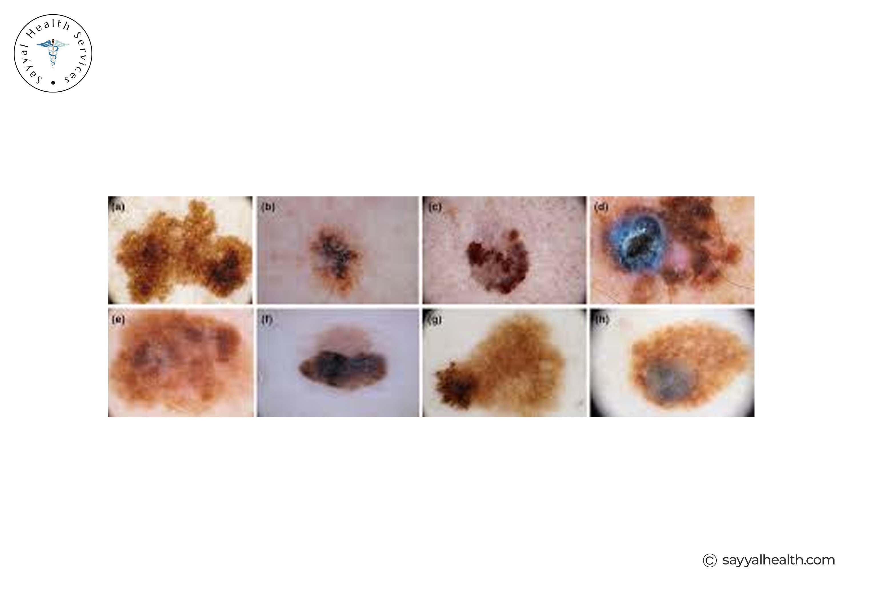
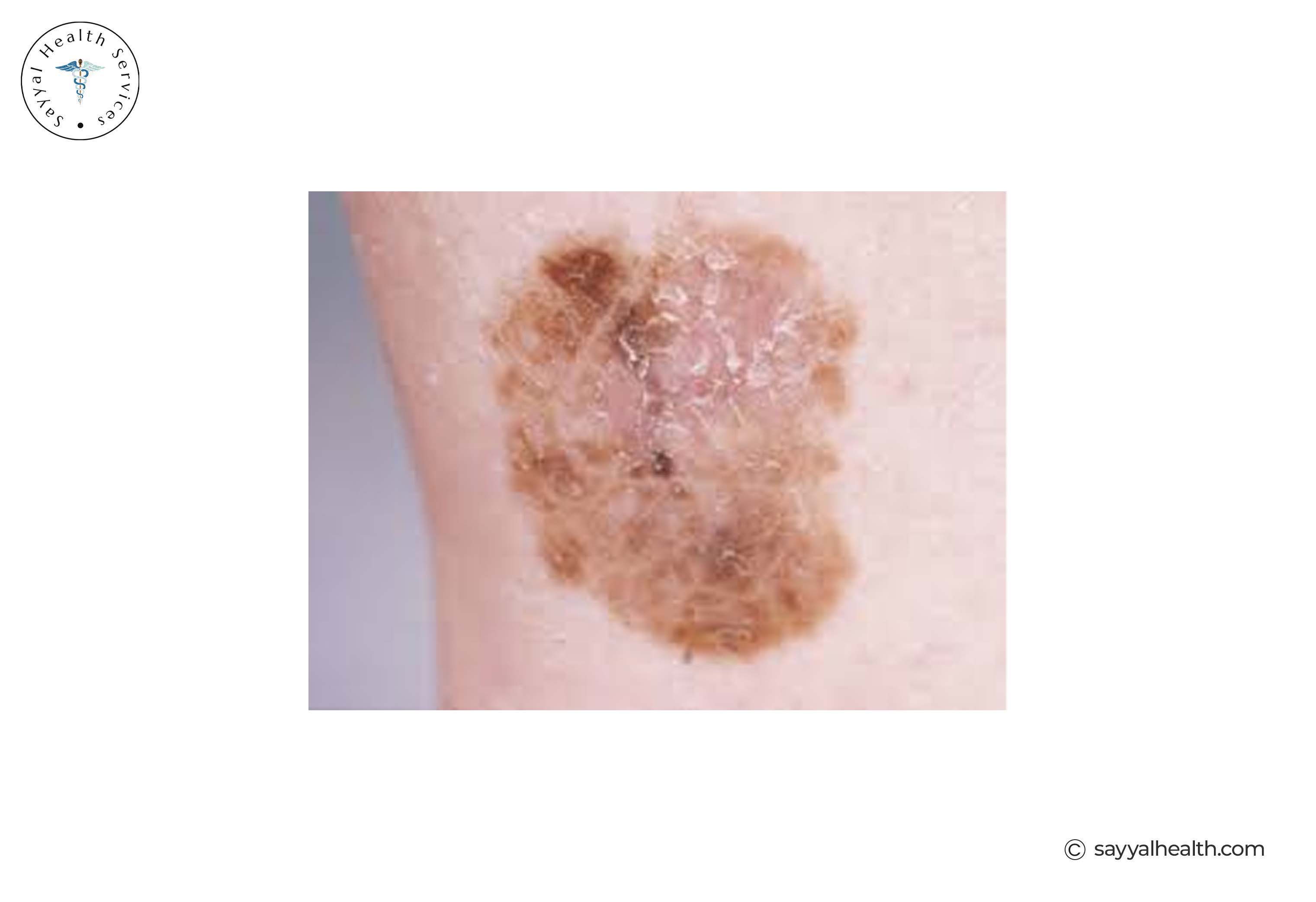
Superficial spreading melanoma is the most common type. It is roughly 50-70% of all melanoma cases.
Appearance: It typically starts as a flat or slightly raised patch with uneven borders and mixed colours including shades of brown, black, pink, red, or white.
Where it appears: Most commonly on the back in men and on the legs in women. It can appear anywhere on the body.
Growth behaviour: It grows horizontally across the surface of the skin before spreading deeper. This is called radial growth. The horizontal phase can last months to years, allowing early detection.
Key difference from other types: Unlike nodular melanoma, it tends to grow outward before growing downward, making early detection more likely if you monitor your skin regularly.
Pictures of superficial spreading melanoma showing irregular borders and mixed pigmentation.
Nodular melanoma is the second most common type. Also, it is one of the most aggressive forms.
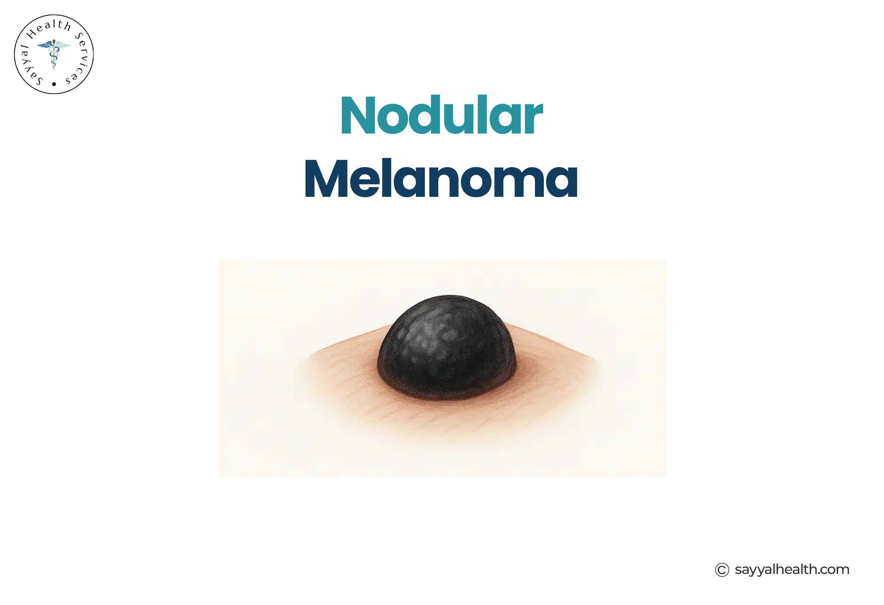
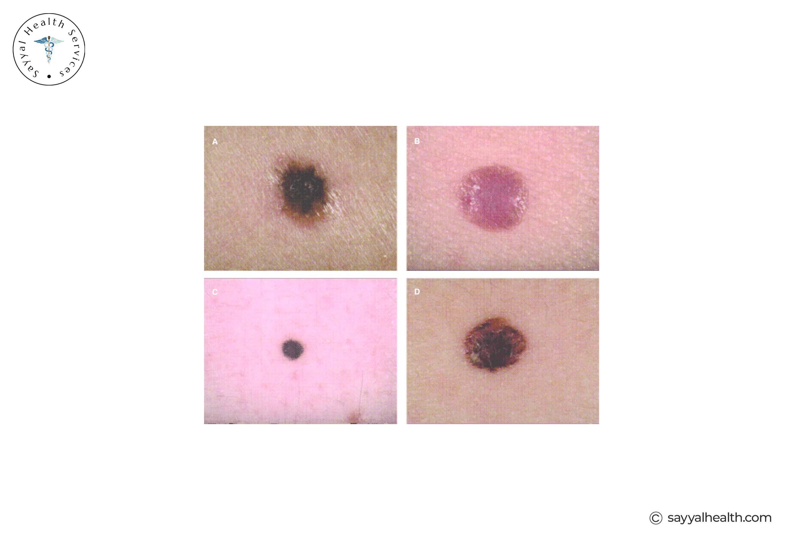
Appearance: It appears as a raised and dome-shaped lump. It is often dark brown or black, but can also be red, pink, or skin-coloured. Nodular melanoma does not go through a prolonged flat phase.
Where it appears: It can develop anywhere on the body. Specifically, nodular melanoma on the scalp is common and often missed due to hair coverage.
Growth behaviour: Unlike other types, nodular melanoma grows vertically from the start, directly into deeper layers of skin. This makes it faster-growing and more dangerous.
Early stage nodular melanoma may look like a new mole or a blood blister. Any new raised lump that changes or bleeds should be assessed urgently.
Risk factors: More common in older adults and men. UV exposure plays a significant role.
Nodular melanoma photos showing a raised dark lesion.
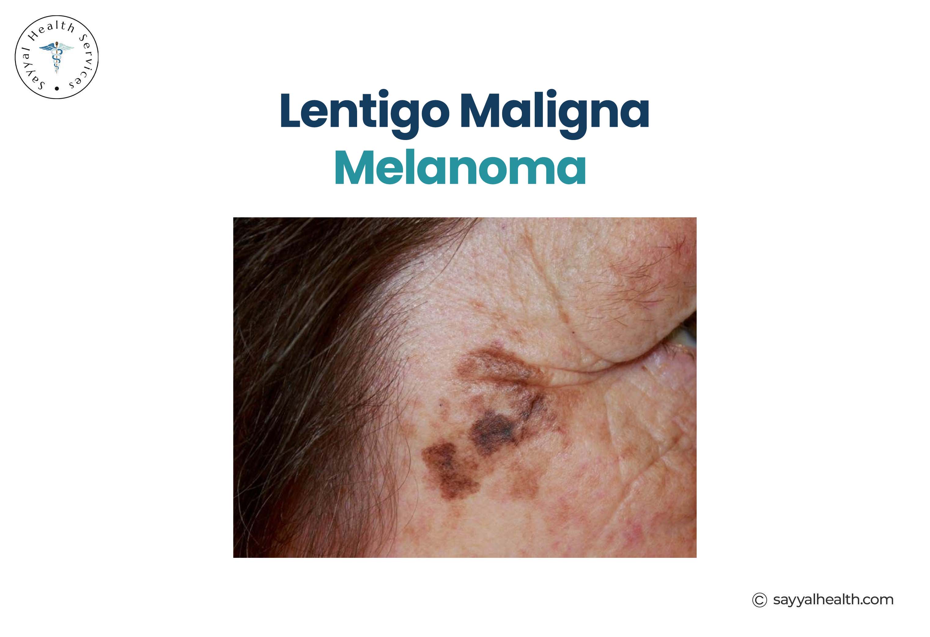
Lentigo Maligna Melanoma begins as a pre-cancerous condition called lentigo maligna. It is a slow-growing flat lesion that eventually develops into invasive melanoma.
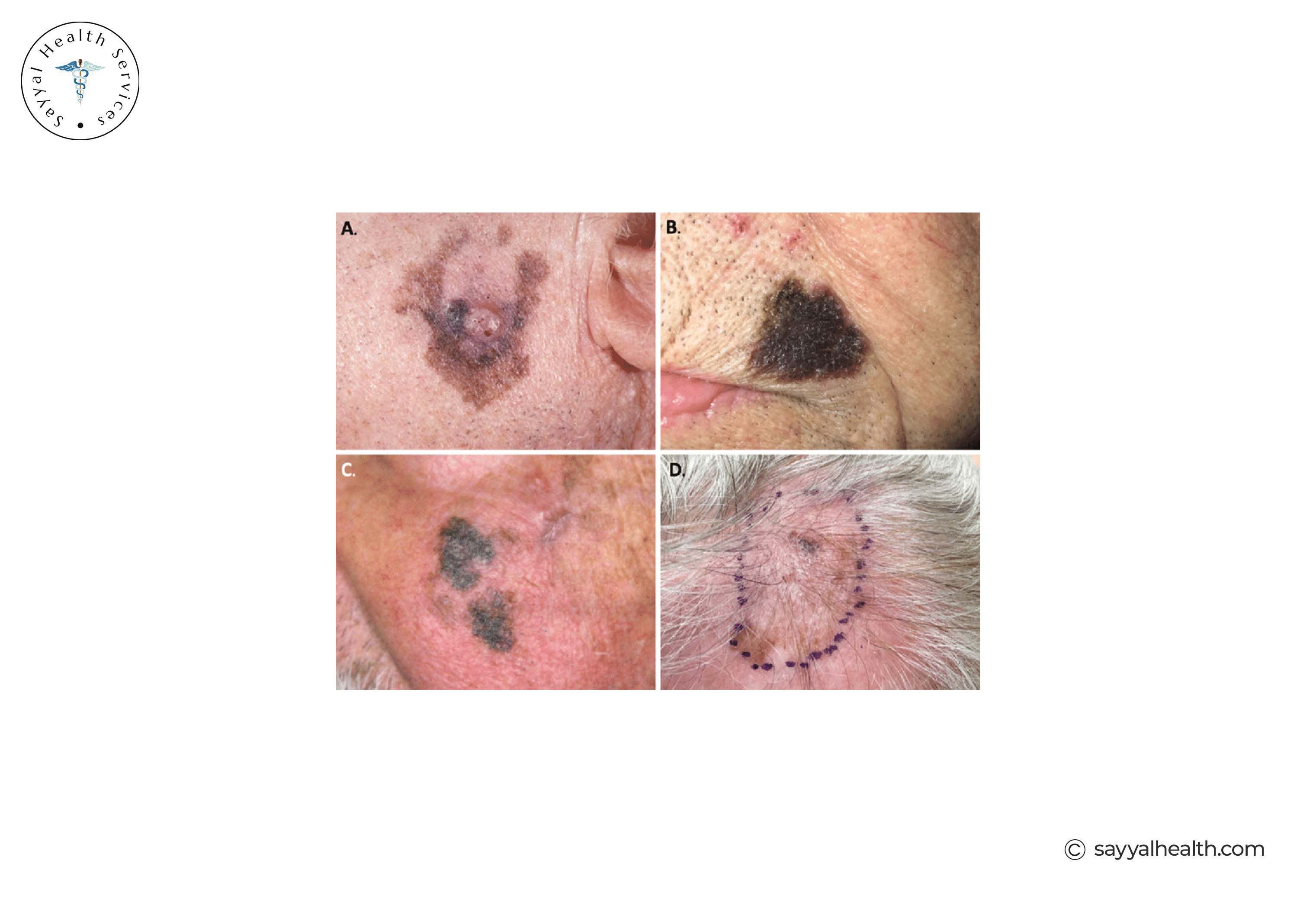
Appearance: A large, flat and irregularly shaped patch with varying shades of brown, tan, and black. It can look similar to an age spot but grows slowly over the years.
Where it appears: Typically on sun-damaged skin in older individuals. The face, nose, ears, and neck are common sites. Melanoma commonly occurs on the face, especially the cheeks and nose.
Growth behaviour: Lentigo maligna can take years or even decades to become invasive. Once it does, it is classified as lentigo maligna melanoma.
Superficial spreading melanoma vs lentigo maligna: Both begin as flat lesions, but lentigo maligna grows far more slowly and is more closely linked to chronic sun exposure in older people. Superficial spreading melanoma tends to appear in younger patients and in more varied body locations.
Pictures of lentigo maligna melanoma on the face.
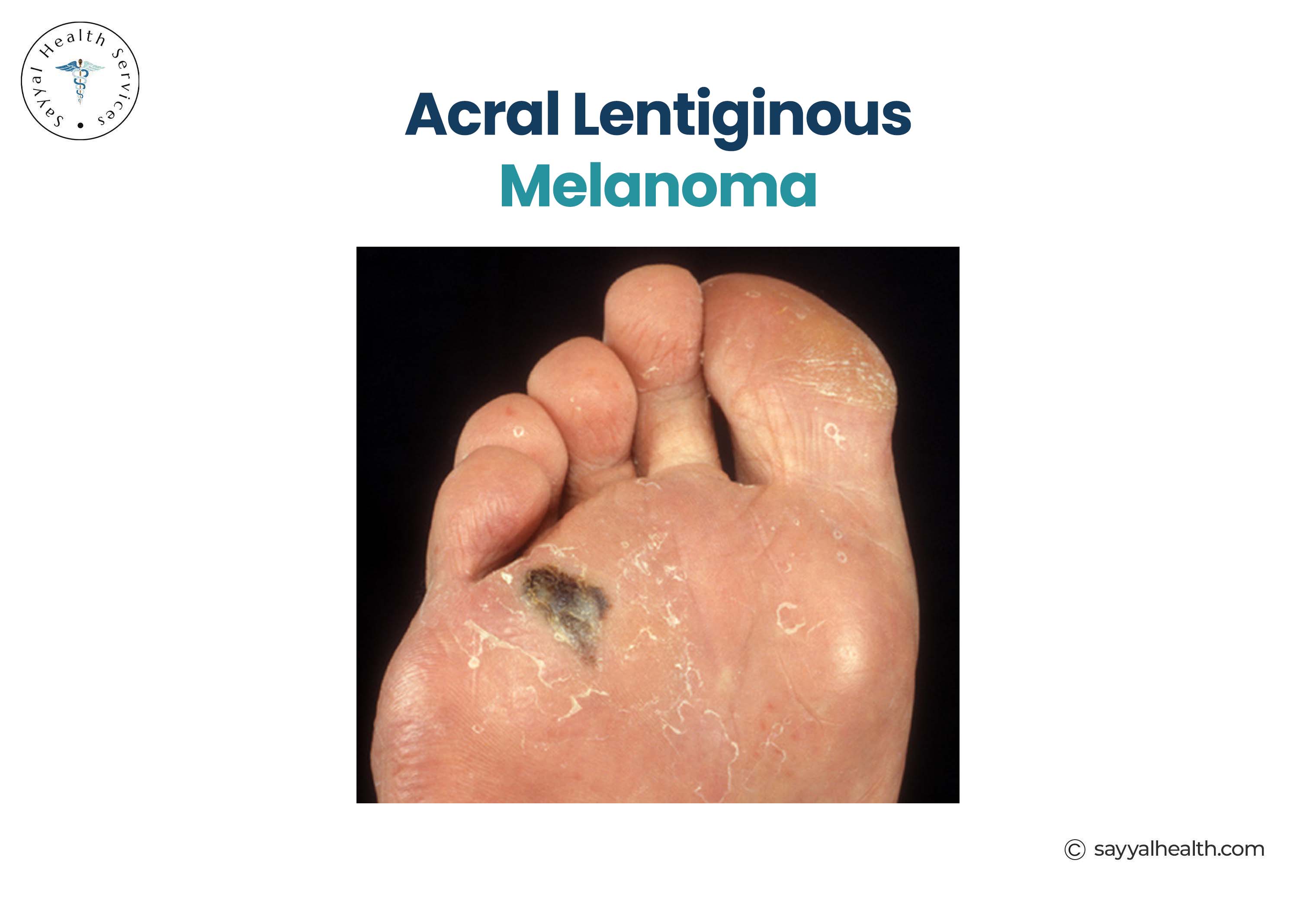
Acral lentiginous melanoma is unique because it appears on areas of the body not typically exposed to the sun.
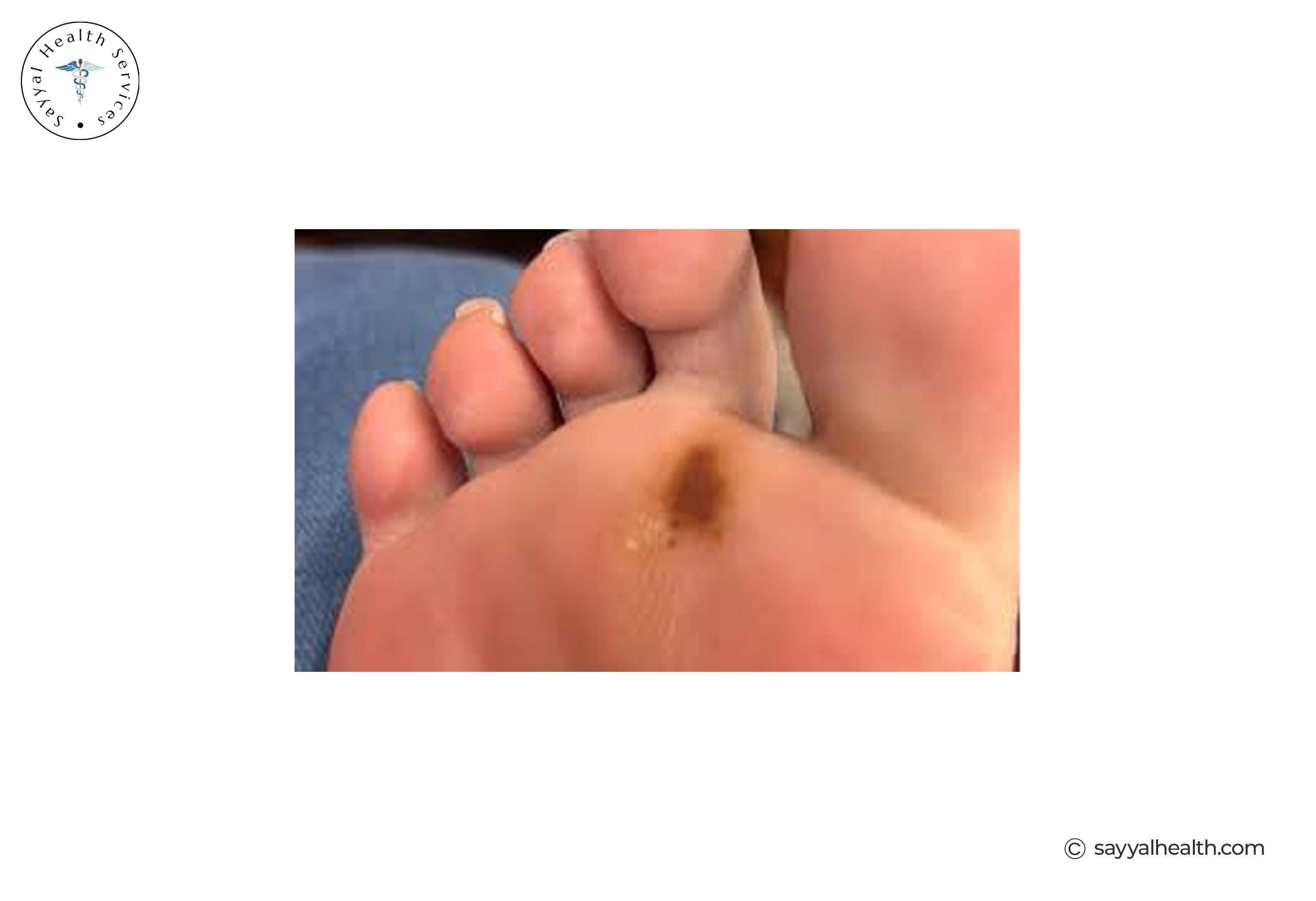
Appearance: A flat, irregularly pigmented patch, usually dark brown or black, that may look like a bruise or stain. Over time, it can become raised.
Where it appears: On the palms of the hands, soles of the feet, and under fingernails or toenails. Skin cancer on the bottom of the foot is most commonly this type. It is also called subungual melanoma when it appears under the nail.
Growth behaviour: It starts in the radial phase, spreading across the surface, then transitions to vertical growth.
Important note: Acral lentiginous melanoma is the most common melanoma type in higher proportion of people with darker skin tones. It appears in less visible locations. That’s why it is often diagnosed late.
Pictures of acral lentiginous melanoma on the sole of the foot.
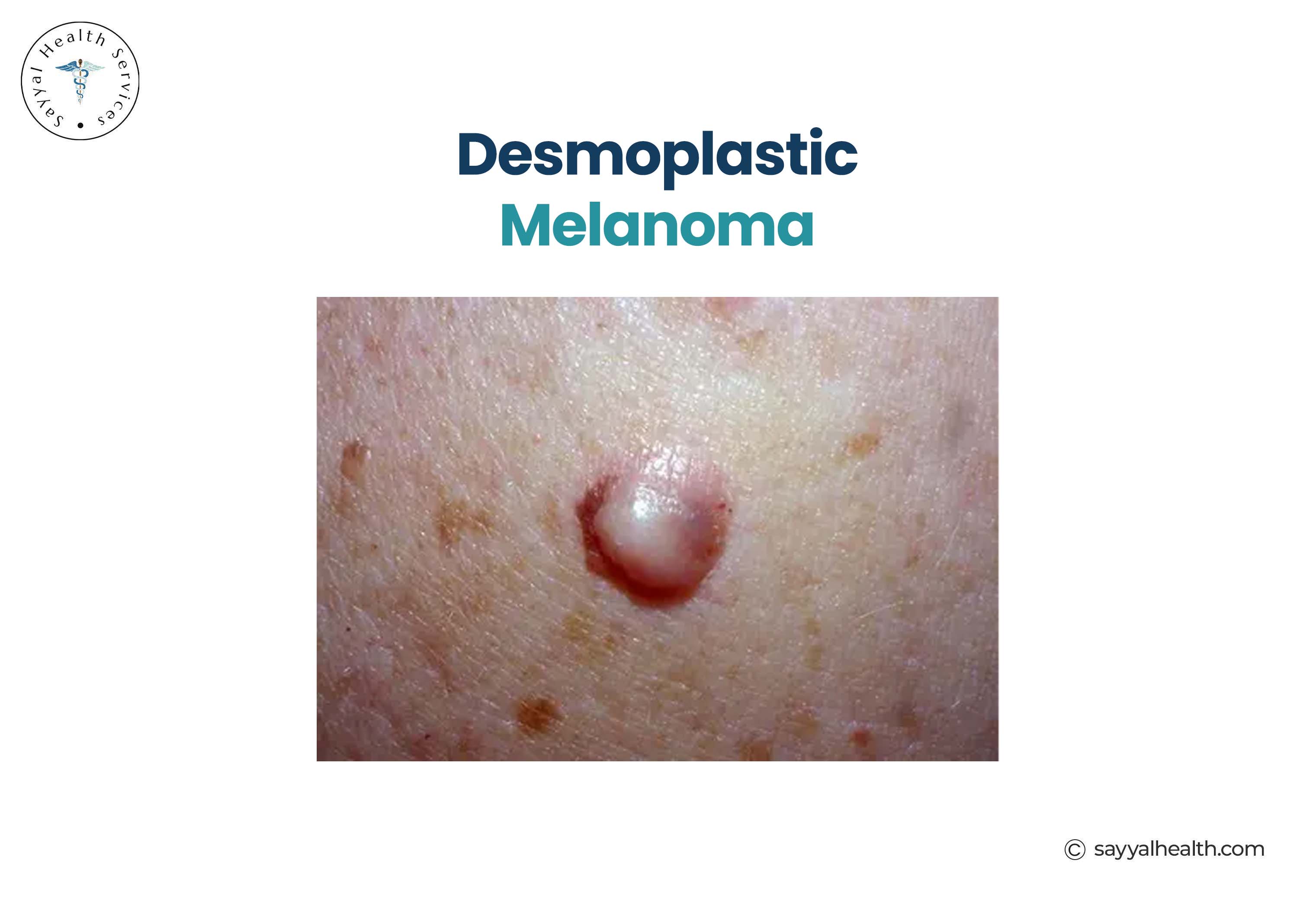
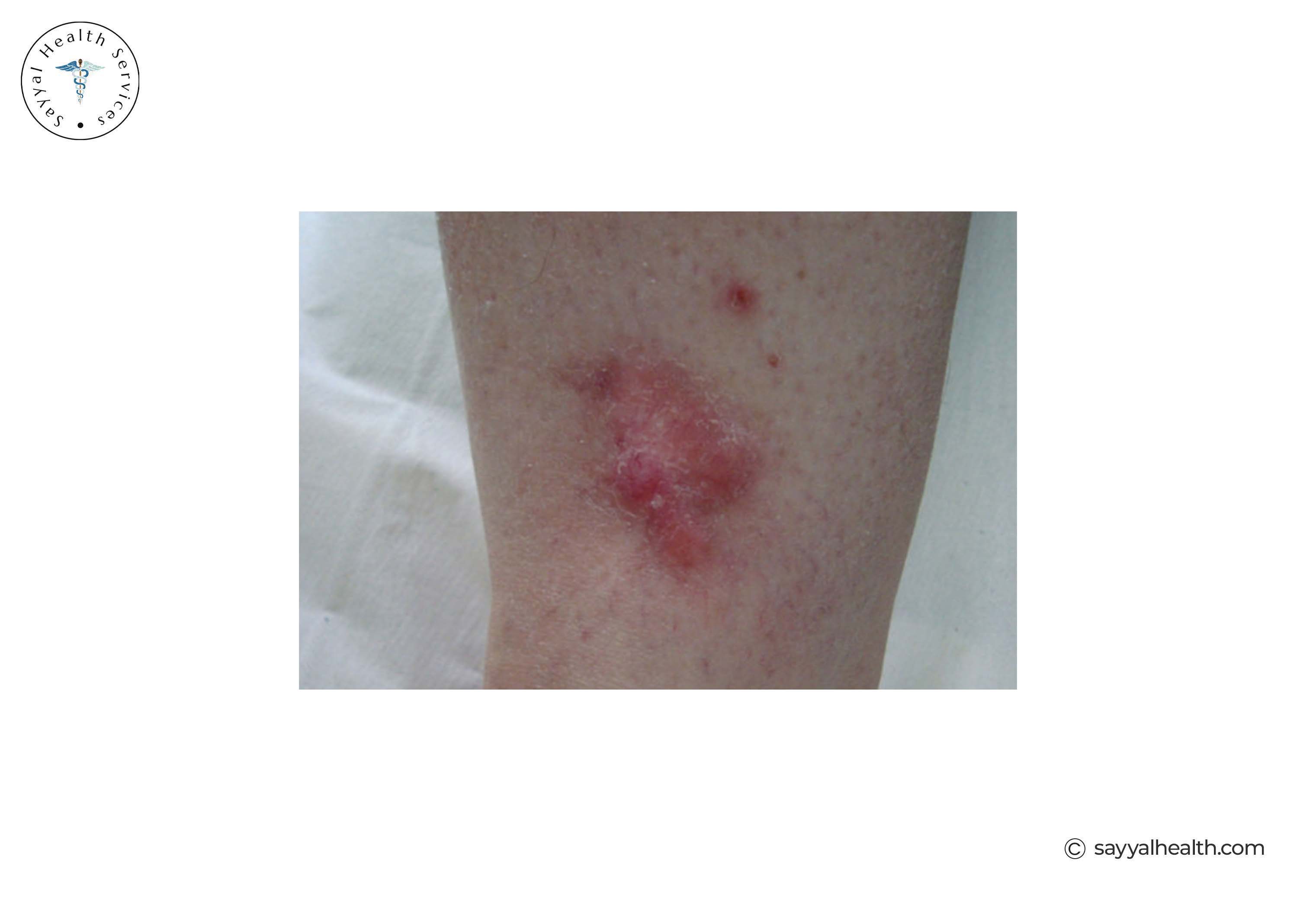
Desmoplastic melanoma is a rare form of melanoma that develops in fibrous tissue around nerve fibres.
Appearance: Often looks like a scar, firm flesh-coloured or lightly pigmented lump. It may not have typical melanoma colours, making it easy to miss.
Where it appears: Commonly on the head and neck, particularly in areas with sun damage.
Key features: It has a high tendency to grow along nerves, making complete surgical removal challenging. Recurrence rates are higher than many other types.
Pictures of desmoplastic melanoma
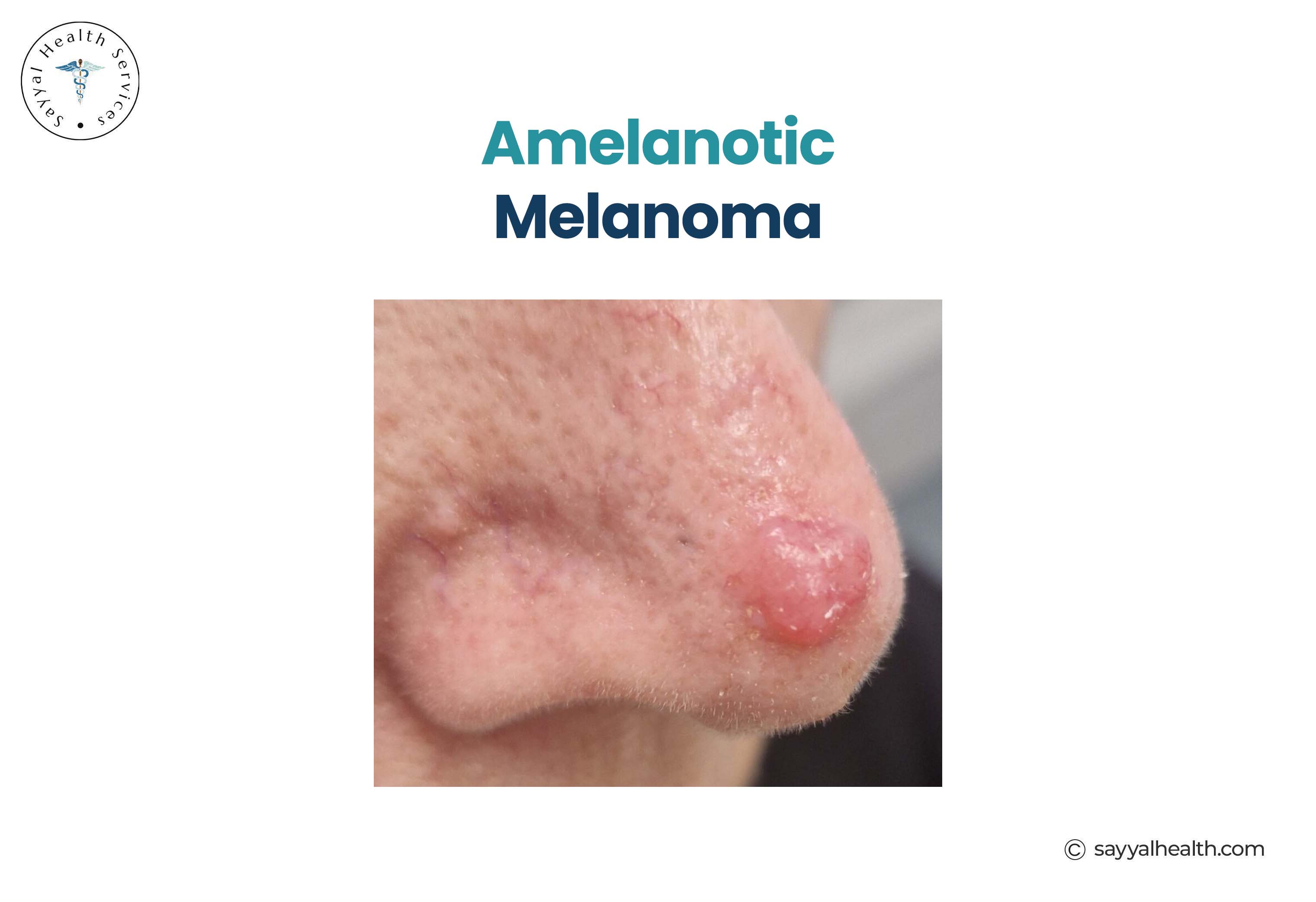
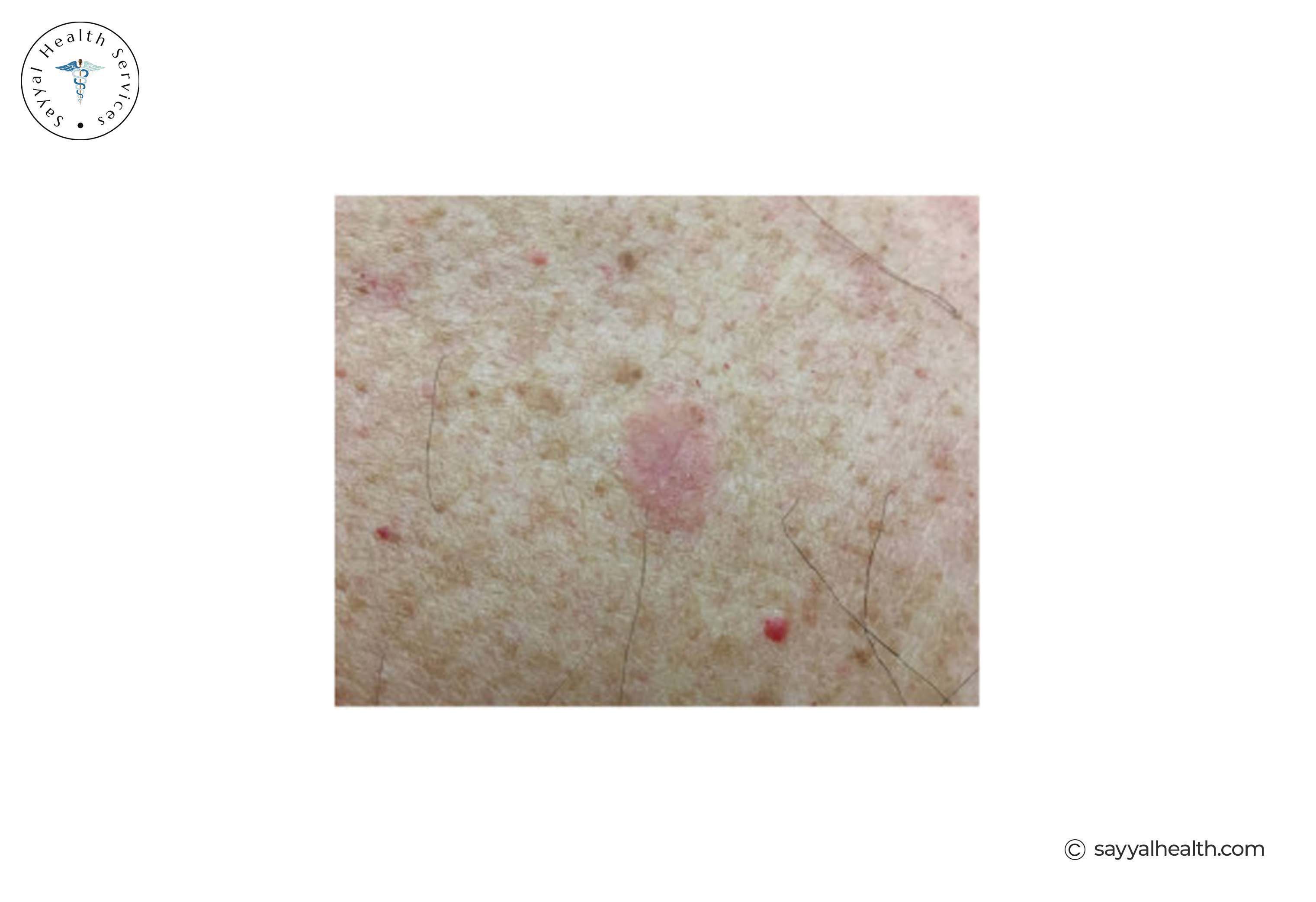
Amelanotic melanoma is a rare and particularly deceptive type because it contains little to no pigment.
Appearance: Pink, red, skin-coloured, or even white. It looks nothing like the typical dark mole most people associate with melanoma. It can resemble a cyst, scar, or benign growth.
Why it is dangerous: Because it lacks the dark pigmentation most people expect, it is frequently misdiagnosed or dismissed. Any persistent pink or red skin lesion that does not heal should be professionally assessed.
Amelanotic Melanoma Picture
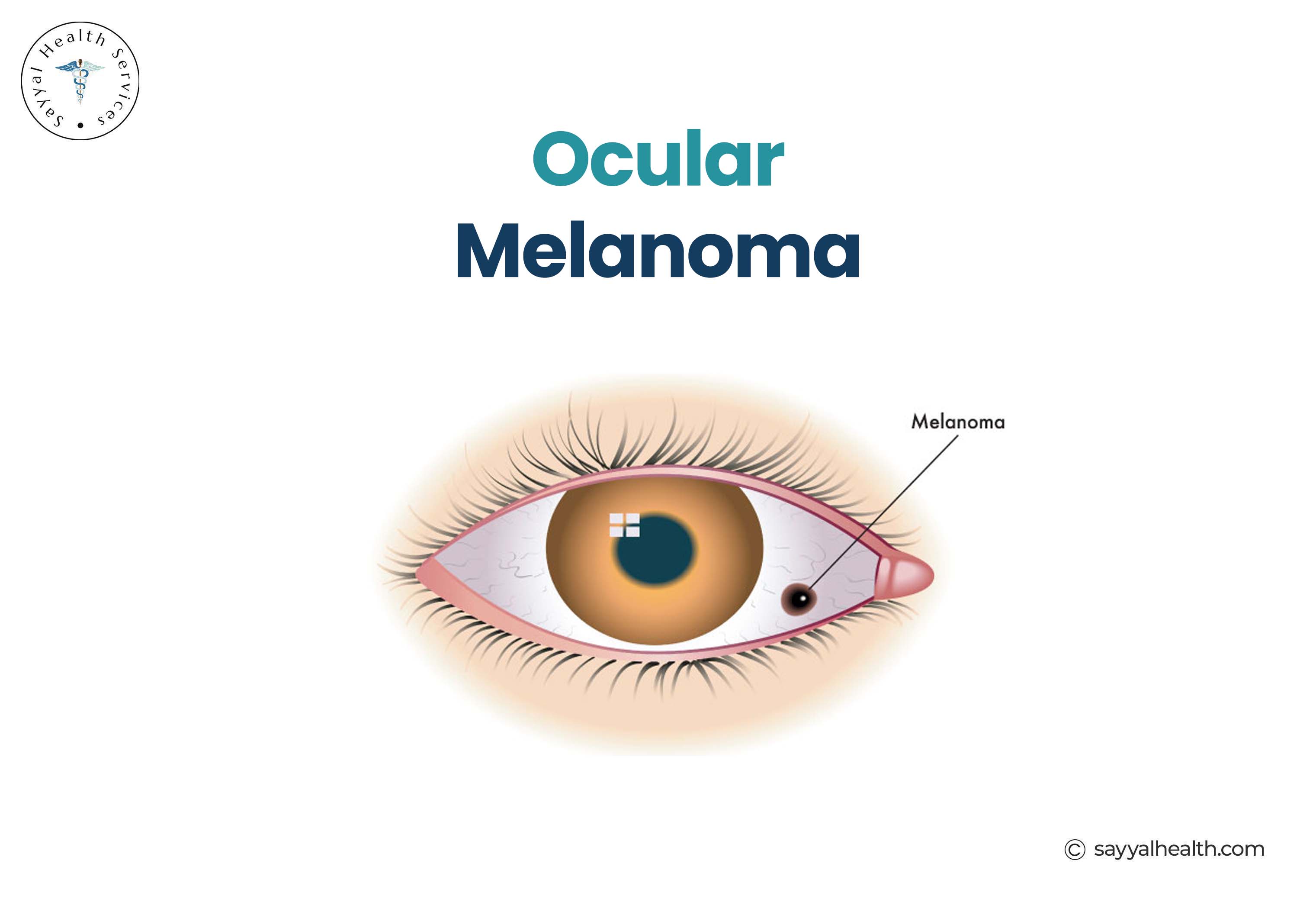
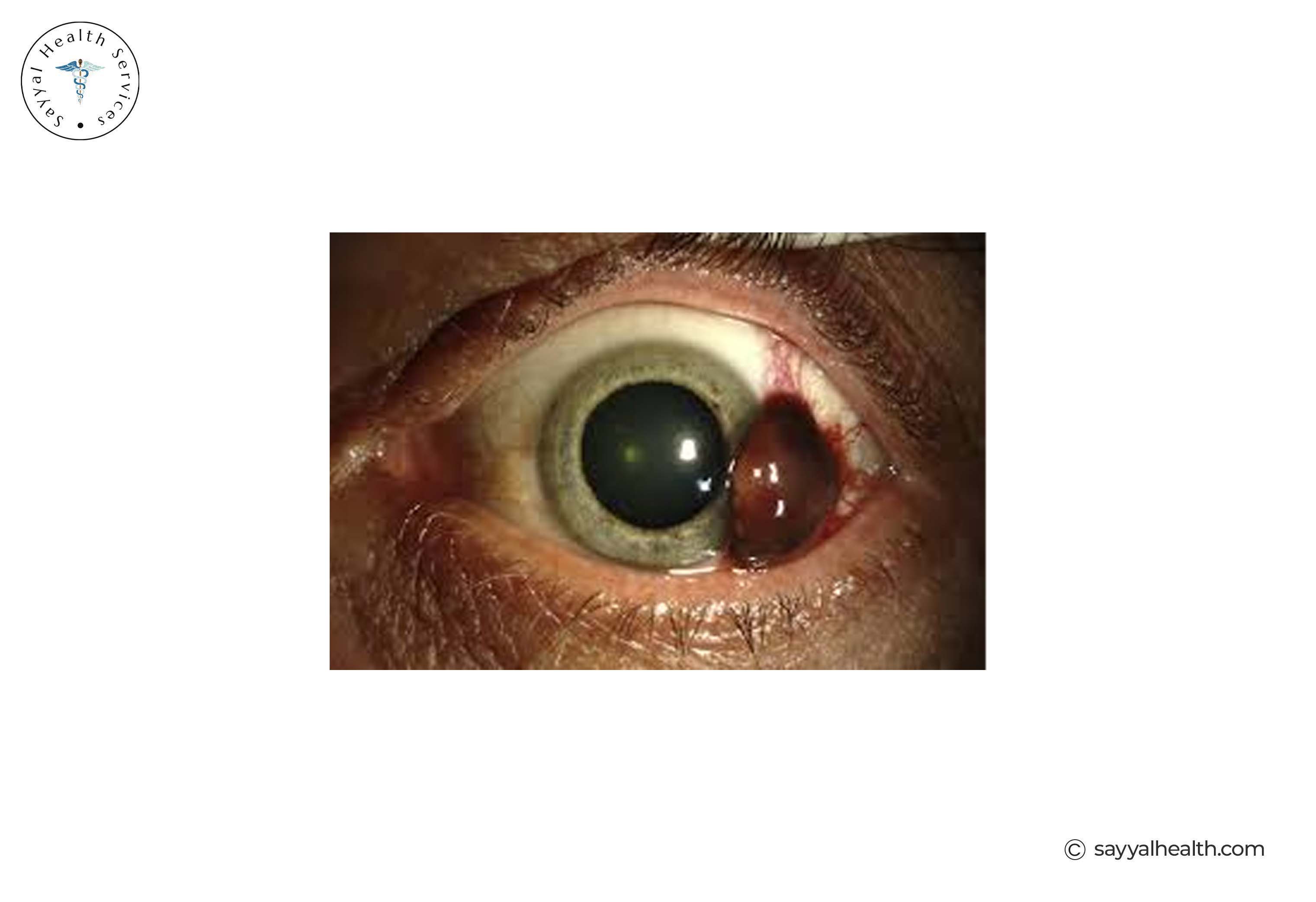
Ocular melanoma, or melanoma of the eye, is the most common primary eye cancer (not skin cancer) in adults. Unlike most melanomas that develop in the skin, ocular melanoma develops in the tissues of the eye.
Where it develops: It usually forms in the uveal tract, the middle layer of the eye. It can also affect the conjunctiva, the surface of the eye.
Symptoms: Many people have no symptoms at all. Some notice blurred vision, floaters, or a change in the shape of the pupil.
Why early detection matters: Ocular melanoma often develops without noticeable symptoms, so it is frequently detected during routine eye examinations.
Photos of Ocular Melanoma
Mucosal melanoma develops on the mucous membranes that line body cavities including the mouth, nasal passages, throat, oesophagus, genitals, and rectum.
Why it is concerning: It is not linked to sun exposure, which makes prevention difficult. It is often diagnosed late due to its hidden location.
Symptoms vary depending on location. Bleeding, discharge, or a visible lesion may be present.
Photo of Mucosal Melanoma
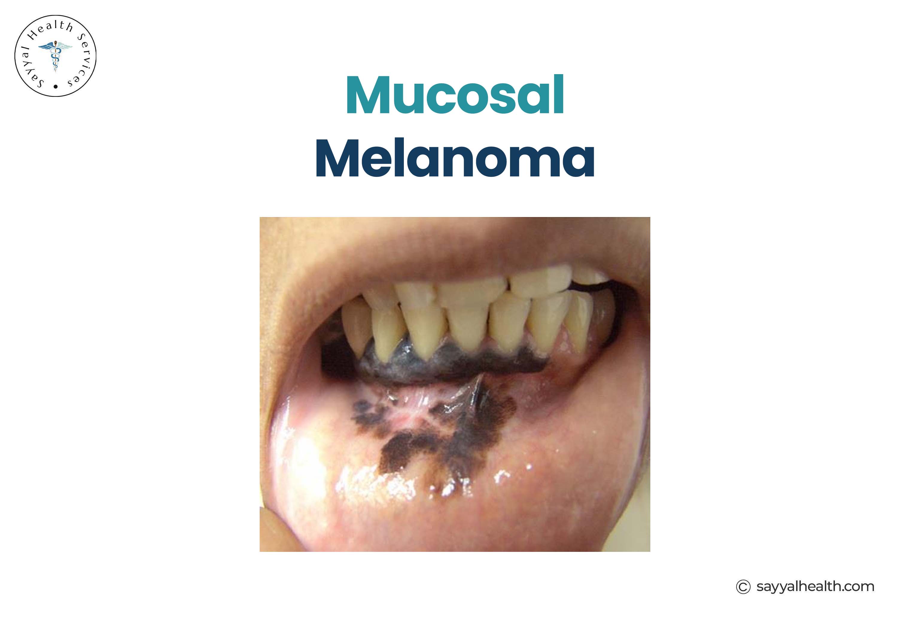
Mucosal melanoma is different from cutaneous melanoma because it develops in the mucous membranes rather than the skin.
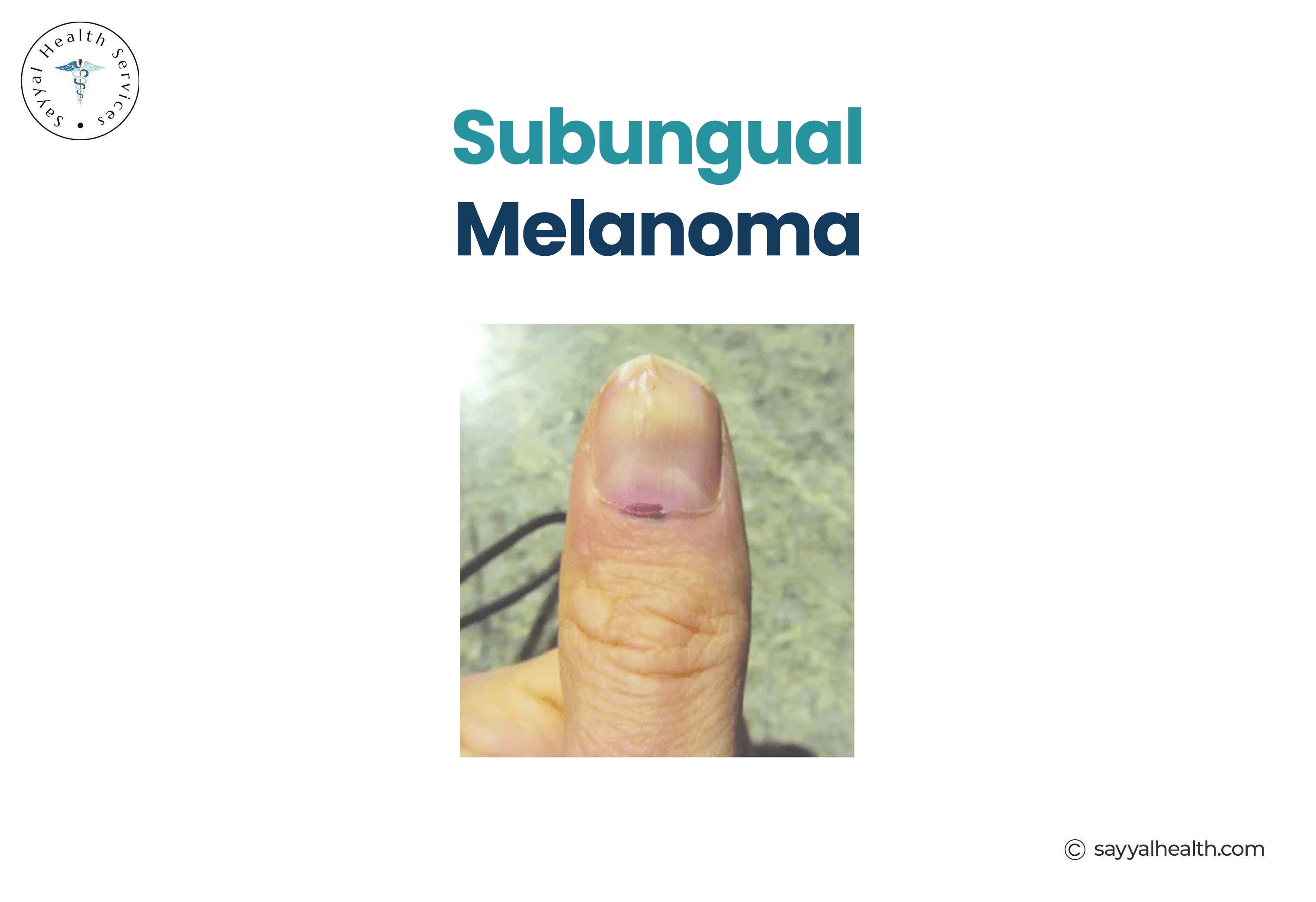
Subungual melanoma is a form of acral lentiginous melanoma that develops under the fingernail or toenail.
Appearance: A dark brown or black streak running lengthwise under the nail. It may look like a bruise that does not move as the nail grows.
Key distinction: A dark band under the nail that persists and widens should always be assessed by a doctor, especially in adults over 50.
Subungual Melanoma Picture
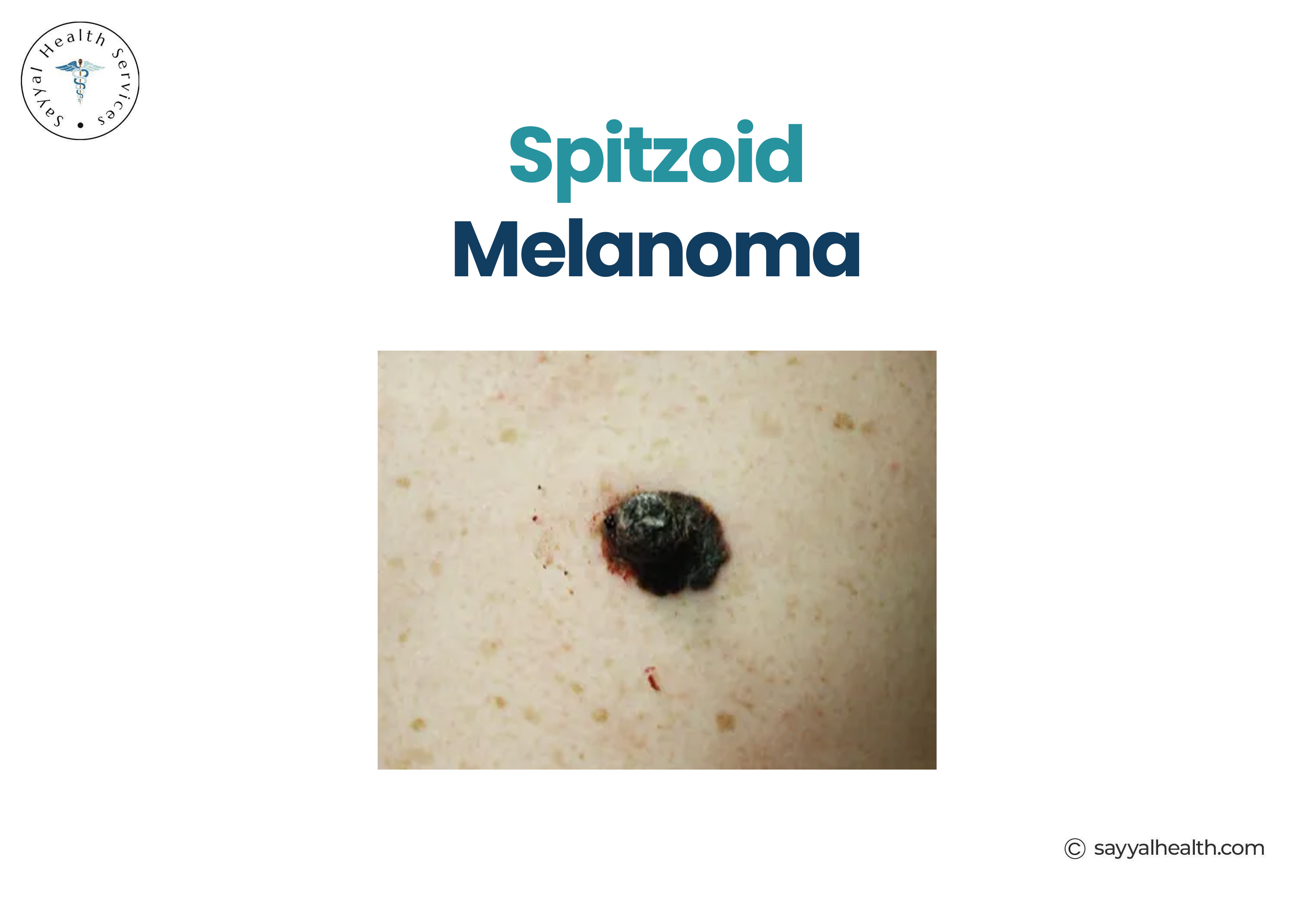
Spitzoid melanoma resembles a Spitz naevus, a type of benign mole, making it one of the most diagnostically challenging melanoma types.
Appearance: Often a pink or reddish raised lesion, most frequently seen in younger patients including children and adolescents.
Why it is difficult to diagnose: Even experienced pathologists can find it hard to distinguish from benign Spitz lesions. Specialist review is often required.
Picture of Spitzoid Melanoma
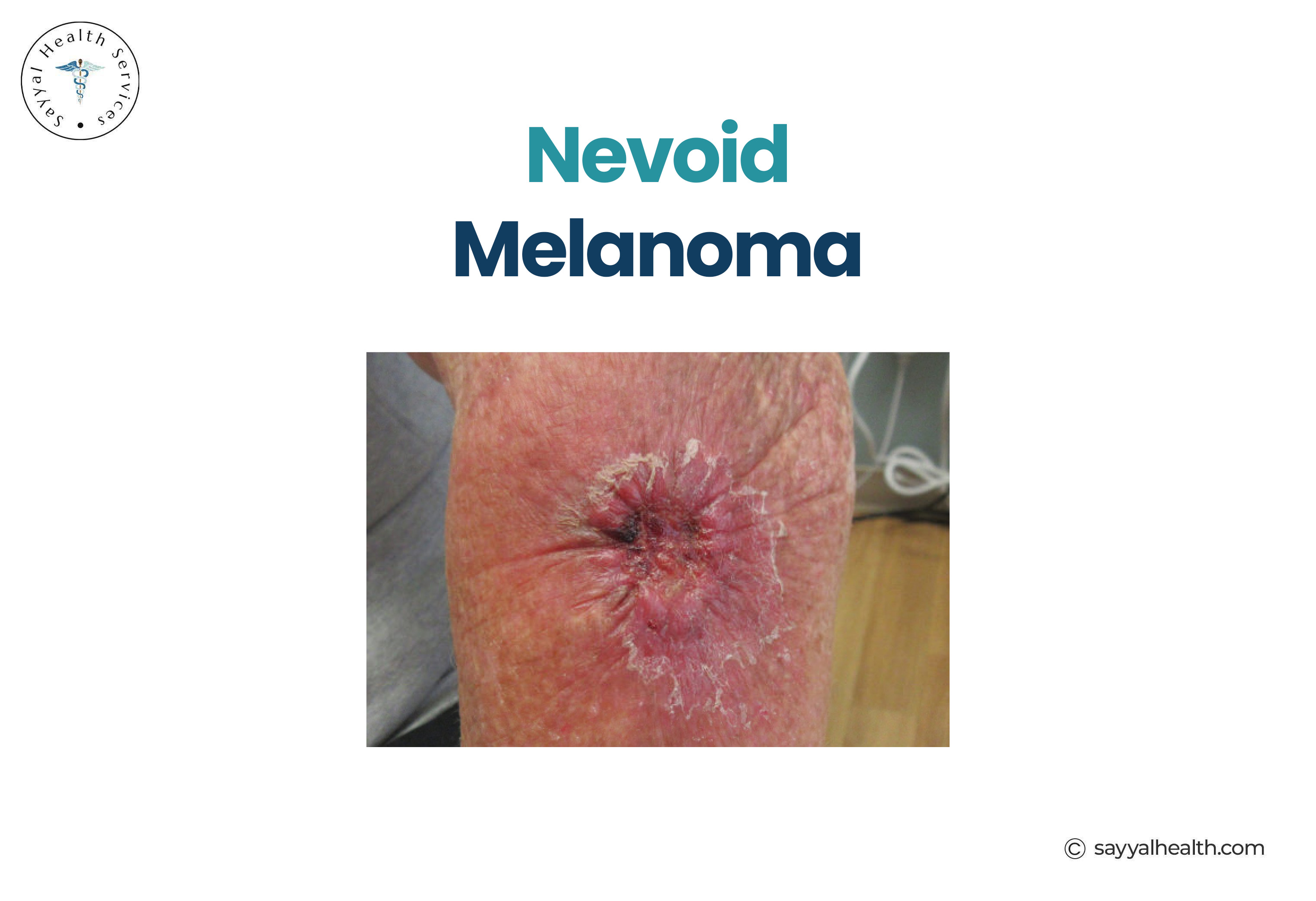
Nevoid melanoma closely mimics an ordinary benign mole in appearance.
Key concern: It can grow slowly and look entirely normal for years, leading to delayed diagnosis. Any mole that changes or feels unusual should be examined professionally.
Nevoid Melanoma Picture
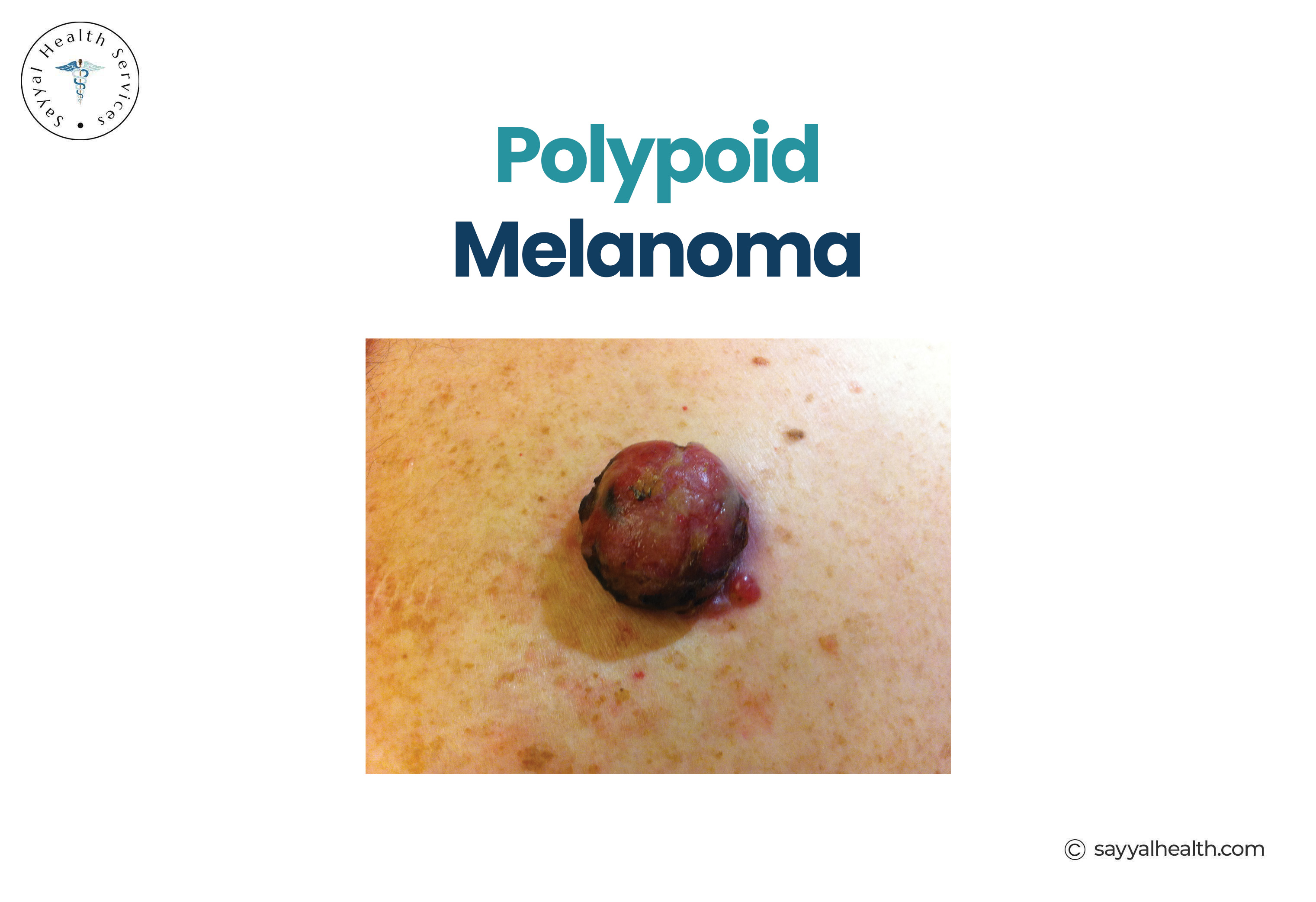
Polypoid melanoma is an aggressive variant similar to nodular melanoma. It grows as a protruding, polyp-like mass and tends to ulcerate and bleed.
Behaviour: Rapid vertical growth and a high risk of early spread to lymph nodes and other organs.
Photo of Polypoid Melanoma
|
Type |
Flat or Raised |
Growth Speed |
Common Location |
|
Superficial Spreading |
Flat, may rise over time |
Moderate |
Back, legs |
|
Nodular |
Raised from the start |
Fast |
Anywhere |
|
Lentigo Maligna |
Flat |
Slow |
Face, Nose |
|
Acral Lentiginous |
Flat, may raise over time |
Moderate |
Palms, soles, nails |
|
Desmoplastic |
Often raised |
Variable |
Head, neck |
|
Amelanotic |
Flat or raised |
Variable |
Anywhere |
|
Subungual |
Under nail |
Moderate |
Fingernails, toenails |
This depends heavily on the type. Nodular melanoma can progress to an advanced stage within weeks to months. Lentigo maligna may remain in its pre-invasive stage for years. Superficial spreading melanoma typically has a slower early phase.
Is melanoma slow-growing? Some types are, but many are not. Even slow-growing types become dangerous once they begin growing vertically into deeper skin layers. Early detection at any stage dramatically improves outcomes.
Protect your skin with early detection and expert care by booking a professional skin checks at Sayyal Health in Gregory Hills.
Australia has one of the highest melanoma rates globally. The key risk factors include:
Sun exposure and UV radiation remain the primary modifiable risk factors.
Melanoma cannot be reliably diagnosed from online images alone. While pictures of melanoma types can raise awareness, only a qualified medical professional can provide a proper diagnosis.
Diagnosis typically involves a clinical skin examination, dermoscopy using a specialised magnifying tool, and a surgical biopsy where tissue is examined under a microscope.
Regular professional skin checks are especially important in Australia, where UV levels are among the highest in the world.
Can melanoma look like a freckle?
Yes, some early melanomas, particularly lentigo maligna, can resemble a freckle or age spot. A melanoma freckle typically grows, changes colour, or develops irregular borders over time.
Can melanoma appear on the foot?
Yes. Melanoma on the foot, often acral lentiginous melanoma, can develop on the sole or between the toes. It is frequently missed because these areas are not routinely examined.
What does amelanotic melanoma look like?
It can look pink, red, or skin-coloured which often resembles a scar or harmless growth. It lacks the dark pigmentation typically associated with melanoma, making it easy to overlook.
Is nodular melanoma always dark?
No. Nodular melanoma can be dark brown or black, but it can also be red, pink, or skin-coloured. Any new raised lesion that changes rapidly should be checked immediately.
Melanoma comes in many forms, and no two types look exactly alike. From the common superficial spreading melanoma to rare types like desmoplastic and mucosal melanoma. Each type has distinct features, growth patterns, and locations.
The most important thing to remember is that early detection saves lives. Melanoma that is caught early is highly treatable. Melanoma caught late is far more difficult to manage.
If you have noticed any new or changing spots on your skin, or if you have not had a professional skin check recently, do not wait. Book Appointment with Sayyal Health in Gregory Hills because Early diagnosis is the single most effective way to improve melanoma outcomes.
Remember: This article is for educational purposes and doesn't replace professional medical advice. Always consult with a healthcare provider about any concerns regarding your health.
References:
https://dermnetnz.org/topics/melanoma
https://www.cancer.org.au/about-cancer/types-of-cancer/melanoma
https://www.aad.org/diseases/skin-cancer/melanoma-overview
Your feedback helps us create better content for everyone.